Sunday, November 29, 2009

Inflammation, cancer, and NF-κB

Cancer isn't a single disease, and there are many possible things that can "cause" even a single type of cancer. However, a lot of things basically work in the same general way to promote cancer. One way that's been long suspected though poorly understood is tangled up in the process of inflammation.

At the very center of this story is a transcription factor called NF-κB. It's perhaps best known for its role in inflammation. But we discussed it in connection with cancer over two years ago, here. Since NF-κB is a transcription factor it affects the expression of many genes. Consequently, it's involved in many other biological phenomena, a few of which have been discussed here, here, and here.

Let's review some of the characteristics shared by most forms of cancer. One frequently cited summary, due to Douglas Hanahan and Robert Weinberg, names six "hallmarks of cancer" – self-sufficiency in growth signals, insensitivity to growth-inhibitory signals, evasion of apoptosis, limitless replicative potential, sustained angiogenesis, and tissue invasion and metastasis. It turns out that inflammation and NF-κB can affect most of these hallmarks, but we'll eventually focus on the role NF-κB plays in inhibition of apoptosis (programmed cell death).

Before we get into details, let's go over the way that cancer typically develops. Each of the hallmarks represents a failure in one or more parts of the machinery of cells to properly regulate the cell's life cycle. The bad thing about cancer, of course, is that a certain group of cells – which might all be descendants of a single aberrant cell – begins to grow and proliferate in an unregulated way. Eventually large numbers of unregulated cells will cease to function as required in the organ where they reside – the brain, the liver, breast tissue, or whatever. Instead they will acquire the ability to migrate and take up residence in places where they continue to proliferate and then disrupt the proper function of whatever organ they wind up in. This is metastasis, and it is usually fatal to the animal in which it occurs.

The cell's machinery is ultimately controlled by the way specific genes of the cell's DNA are expressed to produce proteins. So the reason a cell initially becomes improperly regulated is usually the occurrence of fresh damage to the cell's DNA. It's true that problematic DNA mutations can be inherited from parents, such as the well-known breast cancer genes BRCA1 and BRCA2. Signifcantly, the normal function of both these genes, when not mutated, is to produce proteins whose job is to detect and/or repair DNA damage or to arrest cell proliferation if DNA damage is detected but can't be repaired. So mutated versions of these genes do not exactly "cause" cancer themselves, since some mutation to other genes that actively induce excessive cell proliferation is also required. The potential for cancer may simply remains latent for awhile in cells with mutated genes, regardless of whether the mutation was inherited or occurred much later in life.

But eventually uncorrected DNA damage will occur and affect genes that do induce excessive proliferation. Metastatic cancer eventually results, regardless of whether the first harmful mutations occur in genes that induce proliferation or genes that help regulate proliferation. Cells that contain such mutations divide more frequently and outcompete for resources cells that are functioning properly. So a Darwinian evolutionary scenario arises in which cells that are working "correctly" lose out to cells that become better "adapted" to the job of simply replicating themselves.

The point of this general discussion about cancer is to help clarify what it means to say that something "causes" cancer. In reality, there's never a single event that is the "cause". A number of things have to go wrong before some population of cells with similar defects is numerous enough to outcompete properly functioning cells and to go on to acquire further defects when the mechanisms that normally protect against damage themselves begin to fail.

There appear to be several ways in which NF-κB can contribute to the development of cancer, but one of the least surprising ways is the fact that NF-κB is able to inhibit apoptosis. This ability is not accidental. Most likely it is properly there to enable proliferation of immune system cells in the presence of an infection, as indicated by inflammatory signals.

To return to our main topic, inflammation is a thoroughly normal part of the immune system's operation. NF-κB plays an important part in the inflammatory process. But in simply doing their jobs, these things can, under the right conditions when other cellular mechanisms befome defective, also contribute to the development and progress of cancer.

Normally, the ability of NF-κB to inhibit apoptosis doesn't present a risk of cancer because NF-κB is regulated by other proteins, as we'll discuss later. However, if there are gene mutations that affect NF-κB regulatory proteins, the "safety catch" mechanism may be compromised, and apoptosis may be inhibited when it shouldn't be. Such a problem is more likely to occur when inflammation is present, since then the "safety catch" is already partly disabled.

Another mechanism that keeps NF-κB inhibition of apoptosis in check is the existence of pro-apoptotic proteins such as p53. (We've discussed p53 a number of times before, most recently here, where new research about the anti-cancer properties of p53 is described.) Such proteins are normally subject to regulation themselves, and they become available and active only when needed, such as when DNA damage is detected. However, when one of these proteins has itself been compromised by a gene mutation, inflammation and resultant NF-κB activity can inappropriately inhibit apoptosis, because pro-apoptotic factors are weakened or sidelined.

As it turns out, according to recent research, among the ways that p53 promotes apoptosis is by direct interference with NF-κB's ability to inhibit apoptosis. Further, certain mutations of the p53 gene can remove p53's pro-apoptotic ability, allowing inflammation and NF-κB to contribute to development of cancer. (See here. We'll discuss that in a separate article.)

The high-level view of all this is that cells exist continually in a state of balance between opposing possibilities. Pro-apoptotic and anti-apoptotic mechanisms are not only regulated independently, but they also keep each other in check. But when mutations in any number of possible genes upset the balance, otherwise normal and useful mechanisms can lead to cancer.

Now let's go a little deeper into the subject of various factors that actively contribute to cancer, starting with DNA damage – mutations. The class of things that cause mutations comprises factors such as carcinogenic chemicals, reactive oxygen species ("free radicals"), ultraviolet light, ionizing radiation (e. g. x-rays, radon gas), and some types of viruses. DNA is also at risk of damage every time a cell divides, because mechanisms that copy DNA and verify the copy during division aren't perfect. So old age alone, when DNA has been damaged in various types of cells that have divided too often, can also be a cause of cancer. Dangerous gene mutations can also be inherited, as already noted.

Mutations in genes that code for proteins that directly or indirectly promote cell division are, logically enough, one source of increased cancer risk. A mutation in some gene affecting a constituent of NF-κB would be an example here if, say, the mutation rendered NF-κB less subject to regulation by the proteins that normally regulate it. And as noted, mutations in genes for proteins that detect or repair DNA damage, or simply inhibit cell division when DNA damage exists, would also raise cancer risk. In both cases, the amount of risk also depends on environmental conditions, such as any that could cause inflammation. Mutations in many other genes can also affect cancer risk, when such genes are involved in angiogenesis or cell motility, for example.

Although a wide variety of DNA mutations can raise cancer risk, they may not be sufficient by themselves to actually initiate the development of cancer, because nature has provided cells with many defensive safety mechanisms. On the other hand, mutations aren't always necessary either. There are various external factors that might initiate cancer development even in the absence of DNA damage.

Frequently, it could be an infectious agent like a virus that stimulates excessive cell division. It's quite natural to expect some types of viruses to do this, because such viruses depend on a cell's normal DNA replication machinery to replicate virus DNA as well as cellular DNA. A virus that can easily co-opt the replication machinery has an evolutionary advantage. Especially if the virus can also override normal protective mechanisms, by inhibiting tumor-suppressing proteins like p53. All viruses bring along their own DNA or RNA, which may have evolved specifically because they disable anti-cancer mechanisms. Although virus genetic material is imported from outside, it acts like harmful gene mutations.

Viral replication strategies differ widely among different types of viruses, and certain of these strategies are especially conducive to cancer development. HPV, the human papilloma virus, which is responsible for cervical and anal cancers (among others) is an especially good example. Among the proteins that make up HPV are two, called E6 and E7, each of which promotes cell proliferation in its own way.

E6 is able to suppress the important anti-proliferation protein p53, which manages signals of DNA damage to take appropriate action, such as apoptosis or suspension of the cell cycle. E7 affects the protein pRb, which normally suppresses the cell cycle by binding to a transcription factor known as E2F. When E7 binds to pRb, E2F is released and can go on to advance the cell cycle, which then causes replication of HPV as well as unwanted cell division.

However, what HPV does isn't the only way that an infectious agent such as a virus can promote cancer. Infectious agents also activate the body's immune system to produce an inflammatory response. This inflammation itself can be a cause of cancer. Briefly stated, inflammation causes NF-κB to be activated in order to cause expression of genes that help invoke other immune system components to fight the infection. But NF-κB also has a side-effect of suppressing apoptosis, and as noted above, that is one of the "hallmarks" of cancer.

A connection between inflammation and cancer was suspected over 100 years ago by scientists like Rudolf Virchow. But only rather recently has solid evidence for the connection been found. A good example is Helicobacter pylori bacterial infections associated with stomach cancer (as well as stomach ulcers). A more recent example is an apparent link between inflammation, due to infection caused by the protozoan Trichomonas vaginalis, and prostate cancer. (See here.) Epidemiological evidence suggests that underlying infections and inflammation are associated with 15-20% of all cancer deaths.

There seem to be a number of factors that explain the connection. NF-κB seems to be one of the most important factors, though not the only one. It doesn't work only by suppression of apoptosis either. Promotion of angiogenesis, among other things, also seems to be involved. But most likely we still don't have a very complete understanding of the connection.

It's especially important to understand the connection, because infections aren't the only cause of inflammation. Other suspected causes of inflammation include stress and obesity. Understanding how obesity might promote cancer is obviously of no small importance. There is even evidence that depression may cause inflammation (see here), so that it could also lead to cancer.

In what follows, we're going to encounter various proteins and protein complexes that interact with each other in cell signaling pathways. Often this interaction takes the form that protein A inhibits the activity of protein B; while protein B inhibits the activity of protein C. The net effect is that protein A enhances the activity of protein C, and hence promotes any process that protein C assists in. Or if protein C inhibits some process, protein A will probably do likewise. This complexity can be very confusing, but it's also pretty common, so we just have to deal with it. In fact, the complexity of processes associated with cancer (and much other biology as well) is an important lesson in all of this.

Let's first consider the process of inflammation that occurs "upstream" from NF-κB and activates it. Inflammation refers to the whole process that occurs in a state of hightened immune system activity, due to physiological stress, oxidative stress, infection, or whatever. The outwards signs of inflammation include redness and swelling. There is a beneficial effect of inflammatory activity, of course, in (hopefully) destroying pathogens. But there are harmful side effects as well, including cardiovascular disease, diabetes, a variety of autoimmune diseases, and... cancer.

Many environmental stimuli lead to inflammation and so can cause NF-κB to be activated. Among these stimuli are stress, free radicals, ultraviolet irradiation, oxidized LDL (cholesterol), products of necrotic cell death, and bacterial or viral antigens.

Infectious agents such as bacteria and viruses contain proteins that act as antigens. These antigens are recognized by various cell-surface receptors, especially the kind known as Toll-like receptors (TLRs). Binding by appropriate proteins or antigens ("ligands") to such receptors is sufficient to activate NF-κB, in a manner we'll describe in a moment.

Antigens also bind to and stimulate cells of the immune system to produce various chemical signals such as cytokines, chemokines, and other proteins in order to regulate the immune system response to infection. Among the types of immune system cells that do this are mast cells, dendritic cells, neutrophils, eosinophils, macrophages, and various other lymphocytes. Inflammatory cytokines can also cause activation of NF-κB.

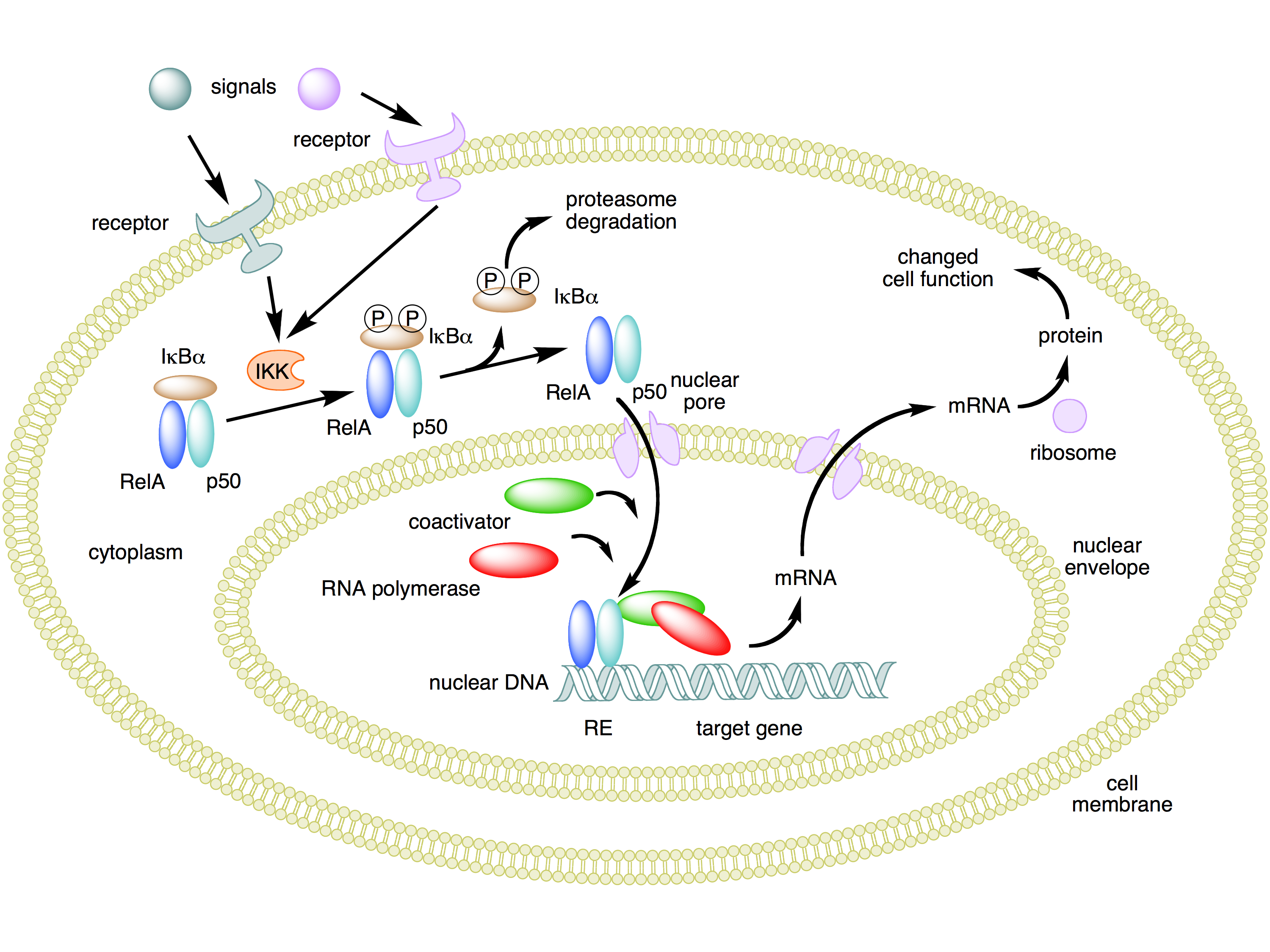
The crucial effect of inflammation for our purposes now is the fact that it activates NF-κB. It's worth noting how this process works. NF-κB is a protein complex consisting of five protein subunits, not a single protein. These subunits are RelA (also known as p65), c-Rel, RelB, p50, and p52. Complexes of these subunits normally circulate outside the cell nucleus, but they are bound to other proteins called IκBs ("inhibitors of NF-κBs") that prevent the complexes from entering the cell nucleus where they could act as transcription factors.

When an inflammatory signal binds to an appropriate cell surface receptor, signals are sent that activate kinases of a family called IKK (IκB kinase). An IKK protein phosphorylates IκBs, which in turn causes them to become unbound from NF-κB and then be destroyed by cellular proteasomes. This frees up NF-κB complexes so that they can enter the cell nucleus and affect the transcription of many genes.

The benefit of an inhibited form of NF-κB existing in the cytoplasm outside the nucleus is that it can be quickly enabled to enter the nucleus and start gene transcription when the need arises. This allows NF-κB to function as a "rapid-acting" transcription factor, without any delays caused by having to wait for the constituent proteins to be synthesized. Here's a diagram that summarizes this process. (Similar considerations apply to p53. The protein is produced and is found in the cytoplasm before it's needed, but kept from activity while bound to another protein, Mdm2.)

The next issue is what happens downstream from the inflammation-initiated activity of NF-κB in the nucleus. Since NF-κB can assist in the transcription of many different genes, the effect of its activation strongly depends on what type of cell it occurs in. Undoubtedly, there's a whole lot we don't yet know about all the affected genes and resulting downstream effects. As far as the immune response – in which NF-κB plays such an important role – is concerned, one effect involves production of cytokines for signaling to other immune system cells.

A second effect is stimulation of cell proliferation. That's generally a good thing if the cell is, for example, a B cell that mediates the part of the immune respose by manufacturing antibodies. But it can also be a bad thing when excessive proliferation of B cells leads to autoimmune diseases, leukemias, or lymphomas. NF-κB stimulates proliferation by enhancing expression of cell cycle proteins like cyclin D1.

However, there's also a third type of effect of NF-κB activity – inhibition of apoptosis. This is especially significant for cancer, because apoptosis is crucial for many natural anti-cancer cellular defenses. In particular, apoptosis is the normal response to severe, uncorrectable DNA damage. It's also the typical way that chemotherapy is able to kill tumor cells. Interference with normal apoptosis makes cancer both more likely to occur, and more difficult to treat.

Exactly how does NF-κB activity inhibit apoptosis? This has been studied, and the answer seems to be that NF-κB inhibits certain enzymes called caspases that are central to apoptosis.

There are even further suspected side effects of NF-κB activity which can play a big role in cancer. One of these is promotion of angiogenesis – production of blood vessels that can supply nutrients to solid tumors. Another is elevated expression of enzymes that promote metastasis.

Observe that the discussion here has been largely theoretical. We haven't described actual experimental research that supports the generalizations. There is some recent research already mentioned that fills this gap. It's especially interesting in the way it exposes a direct connection between NF-κB and p53. But we must leave description of that research for another article, coming very soon.

Further reading:

Nuclear factor-κB in cancer development and progression – May 2006 Nature review article

Tags: ,

Labels: , , ,

Saturday, October 10, 2009

Telomerase and Wnt signaling

Now that research into telomeres and telomerase has (finally) garnered a Nobel Prize, it's a good time to write about recent research on the subject.

Seminal work on telomeres by Elizabeth Blackburn, one of the Nobel winners, was published way back in 1978, and active studies have been going on ever since. So perhaps it's not surprising that the rate of new findings is not so rapid as occurs in newer areas – such as stem cells.

But fascinating new results on telomeres and telomerase do still appear, and one of them connects with more recent research areas – such as Wnt signaling and... stem cells.

Here's the press release:

Discovery pinpoints new connection between cancer cells, stem cells (7/1/09)
A molecule called telomerase, best known for enabling unlimited cell division of stem cells and cancer cells, has a surprising additional role in the expression of genes in an important stem cell regulatory pathway, say researchers at the Stanford University School of Medicine. The unexpected finding may lead to new anticancer therapies and a greater understanding of how adult and embryonic stem cells divide and specialize.

Don't bother getting excited about the "new anticancer therapies" bit. That's just boilerplate that about 77.3% of all press releases dealing with cell biology contain, presumably to impress the rubes. If you need something to get excited about, you might recall that telomerase is also being investigated intensively in connection with issues of aging and longevity, independently from cancer. However, while it's possible that something of medical significance may come from this research, that's probably way down the road.

Before discussing the new research, let's review some of the background on telomeres, telomerase, and Wnt signaling.

To begin with, a telomere is a series of short repeated segments of DNA found at the ends of chromosomes in all eukaryotic cells. In cells of vertebrate animals the repeated segment is TTAGGG (where the letters represent nucleobases: T=thymine, A=adenine, G=guanine).

The total number of nucleotides in these repeated segments varies a great deal from species to species, but in humans it is (initially) about 10,000 nucleotides (or ~1700 complete segments). I say "initially", because part of the telomere is lost every time a cell divides – perhaps 50 to 200 nucleotides per division. Obviously, this means that an adult cell newly derived from a stem cell can divide at most 50-200 times before the telomere is all gone. In practice, the number is a lot less than the maximum, perhaps 40 to 60 times, which is called the Hayflick limit. Cells are programmed to stop dividing on reaching this limit, since otherwise useful DNA would be lost or damaged upon further division.

Why does this loss occur? It seems to be somewhat of an accident of the nitty-gritty details of how DNA replication occurs during cell division. I won't go into that, since it's best explained with some diagrams; you can read about it at Wikipedia. In fact, in the early days of molecular biology (around 1972), what happens at the end of chromosomes during DNA replication was rather puzzling, and the puzzle was called the "end replication problem". Now it's pretty well understood, though somewhat messy.

In any case, the loss of DNA from the telomeres during cell division is a fact, and it conveniently explains the existence of the Hayflick limit, which had been recognized since 1965. What happens when the teleomere is all used up and the limit is reached? Cells that have reached the limit don't necessarily die (though they might), but they do stop dividing, and they enter a static phase of cell life known as senescence.

If you think about it, senescence can be a problem, especially in certain tissues that need to continually replenish their cells, such as skin and the lining of the intestines, as well as hair, fingernails, etc. How is senescence circumvented in such tissues? The answer is (adult) stem cells. It turns out that stem cells are not subject to the Hayflick limit. It's not clear whether they are subject to limits at all.

Like any other cell type, stem cells also divide (by the process technically known as mitosis). But there is one difference from the way "ordinary" cells divide. Stem cells can divide asymmetrically, where one daughter cell is another stem cell, but the other daughter is a "progenitor cell", which is close to a normal, fully-differentiated adult cell of a fixed tissue type. Progenitor cells differentiate further into the final form when they divide, and they are able to divide only a limited number of times.

So how is it that stem cells are able to escape the Hayflick limit, dividing an indefinite number of times, even though they are also subject to the same loss of telomere nucleotides with each division? The answer is the enzyme telomerase, which is able to rebuild shortened teleomeres. It's fortunate for the longevity of complex organisms that telomerase exists, otherwise stem cells would not be able to divide often enough to allow tissues exposed to harsh conditions (such as skin and intestinal lining) to be replenished.

In order to explain the results of the research to be described here we need to say more about telomerase, but first let's summarize the function of telomeres. At first it may seem that all they do is compensate for the sloppy way that DNA replication works, by providing long stretches of expendable DNA at the ends of chromosomes. But there is one other notable function: teleomeres are a useful "cap" at the end of a chromosome that is recognizable to cellular mechanisms responsible for detecting DNA damage. If it weren't for the telomeres, the ends of chromosomes would be indistinguishable from DNA damage, which can result from errors in the replication process, as well as damage due to external agents such as ionizing radiation or harmful chemicals. Cells have various mechanisms for repairing many kinds of damage, but even if repair isn't possible, other mechanisms exist that recognize the damage and prevent further cell division or cause programmed cell death (apoptosis).

Not all DNA damage can be repaired or compensated for by apoptosis or cessation of cell division. Unrepaired DNA damage (however it occurs) is the main cause of cancer (though not the only one). So the existence of the Hayflick limit as a result of teleomere shortening acts as one defense against cancer. Cancer can be defined as the uncontrolled proliferation of cells as a result of DNA damage (affecting existing mechanisms that normally control proliferation) or other causes. So fixed limits on the number of times a cell can divide is one of a number of mechanisms organisms have to guard against cancer.

Before moving on, here's a quick summary of the functions served by telomeres: (1) compensate for the chromosome "end replication problem"; (2) make it possible for cells to distinguish chromosome ends from damaged chromosomes; (3) provide natural limits to the number of times ordinary cells can divide, as protection against cancer.

As noted above, the third of these functions is a problem for stem cells that do need the ability to divide an indefinite number of times. Repair of tissues exposed to harsh conditions is not the only circumstance this ability is needed. Another very important case is that of embryonic development. Multicellular, sexually-reproducing organisms start from a single cell (zygote). Yet there are close to 1014 cells in an adult human.

It is true that a single cell that underwent 47 cycles of cell division could theoretically produce that many cells. But that's really pushing the limits, since some cell types are needed in much larger numbers than others. The bottom line is that embryonic development is the other main circumstance when limits on cell division need to be overcome.

Telomerase is what makes this overcoming of telomere limits possible. And it does it in a pretty straightforward way. Telomerase is a complex molecule with three distinct parts. Two of these are proteins: Telomerase Reverse Transcriptase (TERT) and dyskernin, which are coded for by distinct genes. TERT does most of the work. The other part is a short piece of RNA, called the telomerase RNA component (TERC), which contains and is somewhat longer than the repeat unit (TTAGGG in vertebrates). Like any other reverse transcriptase enzyme (other examples of which occur in RNA viruses such as HIV), TERT simply translates a piece of RNA into DNA and inserts it into a chromosome. In the case of telomerase, the RNA is carried inside the enzyme complex itself. Telomerase does its job simply by replacing the telomere DNA that is lost from chromosomes during mitosis.

So telomerase performs the function of rebuilding telomeres, and this is essential in tissues where cells must proliferate rapidly, such as in embryonic development and tissue regeneration. But as we noted above, if the restriction on cell division is circumvented, the risk of cancer goes up. Although telomerase serves an essential purpose in specialized contexts, it also makes it possible for cells to become cancerous. Consequently, telomerase is not expressed in most adult body cells. However, telomerase is expressed in about 90% of tumor cells.

It might seem as though one approach to controlling or even destroying cancer could involve either inhibiting telomerase or developing a vaccine using telomerase to induce an immune response against telomerase-rich cancer cells. There are in fact various clinical trials exploring both techniques. But this is tricky and rather risky, because as we've observed, telomerase is needed in stem cells required for tissue regneration, at least once the cells have begun proliferating. Those cells need to be protected while they are simply doing their job – replenishing skin and intestinal linings, for example. We need to understand how such cells are controlled so that they work without leading to cancer.

Anyhow, TERT is a protein that is an essential component of telomerase, which plays an important role in cell proliferation. The new research we're finally almost ready to discuss shows that, surprisingly, TERT also plays a role in a completely different aspect of cell proliferation having nothing to do with telomeres. That's where Wnt signaling comes in.

Wnt signaling is a subject we've looked at several times before. Some of the relevant articles are here, here, here, and here. Wnt was first noticed in connection with embryonic development and tissue regeneration. This article has many examples. The name Wnt alludes to a gene called "wingless", because the gene causes fruit flies to lack wings when the gene is mutated.

Recently Wnt's importance in stem cells has also received a lot of attention. Some of our discussion of that may be found here, here, here, and here. Wnt's relevance to cancer and cancer stem cells is often touched on in most of all the articles listed.

Basically, Wnt is the name of a family of proteins that play an important role in signals promoting cell proliferation, especially in the context of embryonic development and tissue renewal (skin, intestines, hair, and immune system cells). Since Wnt proteins promote proliferation, they also play a role in cancer.

The fact, then, that new research shows teleomerase can enhance Wnt signaling is signficant as a second, entirely separate route through which it plays a role in both normal stem cell function and cancer.

Here's the research abstract:

Telomerase modulates Wnt signalling by association with target gene chromatin (7/2/09)
Stem cells are controlled, in part, by genetic pathways frequently dysregulated during human tumorigenesis. Either stimulation of Wnt/β-catenin signalling or overexpression of telomerase is sufficient to activate quiescent epidermal stem cells in vivo, although the mechanisms by which telomerase exerts these effects are not understood. Here we show that telomerase directly modulates Wnt/β-catenin signalling by serving as a cofactor in a β-catenin transcriptional complex. The telomerase protein component TERT (telomerase reverse transcriptase) interacts with BRG1 (also called SMARCA4), a SWI/SNF-related chromatin remodelling protein, and activates Wnt-dependent reporters in cultured cells and in vivo. TERT serves an essential role in formation of the anterior–posterior axis in Xenopus laevis embryos, and this defect in Wnt signalling manifests as homeotic transformations in the vertebrae of Tert-/- mice. Chromatin immunoprecipitation of the endogenous TERT protein from mouse gastrointestinal tract shows that TERT physically occupies gene promoters of Wnt-dependent genes. These data reveal an unanticipated role for telomerase as a transcriptional modulator of the Wnt/β-catenin signalling pathway.

That's a pretty good summary of the paper, but I imagine most people would like a bit more explanation of what's going on.

Previous research had disclosed some interesting "coincidences" involving stems cells and embryonic development, in which both telomerase and Wnt signaling seemed to have similar effects, even though no obvious connection was known. For example, epidermal (skin) stem cells spend most of their time in a quiescent (non-dividing) state. The main function of Wnt proteins is to carry signals between cells that inform target cells of a need to start dividing, for example during various stages of embryonic development or to heal wounds. The curious thing is that telomerase was known to have a similar effect as Wnt signaling on some stem cells. This coincidence is enough to motivate looking for a connection.

If you are really into this sort of thing, you might want to refer to this diagram of various cell signaling pathways, including that of Wnt. One of the things it illustrates is the position of β-catenin in the Wnt pathway. β-catenin operates at the end of the pathway, where it becomes a part of a protein complex that includes transcription factors (known as TCF/LEF), and the complex causes expression of a variety of Wnt-regulated genes, which go on to enable cell proliferation.

One of the main findings of the research is that TERT interacts with another protein called BRG1 (or SMARCA4), and together these become important parts ("cofactors") of the gene-regulating protein complex. It was also determined that TERT is the only component of telomerase that is involved with Wnt signaling.

Apparently TERT is more important for some instances of Wnt signaling than for others. For example, low levels of TERT in embryos of the frog Xenopus laevis resulted in very abnormal development of the frog embryos (in terms for the anterior-posterior axis of the body). But low levels of TERT at a later stage of development caused only somewhat more subtle defects in formation of ribs in the embryo. Similar somewhat minor effects also occured with low levels of TERT in mouse embryos, and these defects were much like the effects of low levels of β-catenin.

So what, then, is so interesting about this research? It's satisfying to know the reasons for what were previously just observed coincidences, such as the fact that telomerase is not only very important for the ability of stem cells to divide freely (during embryonic development and tissue regeneration) but that it also can help stimulate stem cell division.

But what makes this research especially significant is the importance of the biological processes in which both telomerase and Wnt signaling play major roles – namely embryonic development, tissue regeneration and repair, and cancer. The latter two processes are especially important for medical reasons, although we're still a long way from being able to use this knowledge about telomerase and Wnt signaling for therapeutic purposes.

As far as cancer is concerned, we need to understand much more about how telomerase and Wnt signaling work in various types of stem cells, given that stem cells may play a big role in some types of cancer (though not in others).

But tissue regeneration and repair are also of significant medical interest, since it is the eventual inability of various types of tissues to replenish themselves in old age that is responsible for the many debilities that appear in old age. Many people have speculated that telomerase could help alleviate this problem – provided it does not also lead to facilitating the development of cancer.

Finally, we are left with the puzzle of how it is that TERT happens to play important – yet quite distinct – roles in two very separate processes that are, neverthelesss, both important for cell proliferation. We don't know the answer to this, but we can speculate. Perhaps it's because TERT has to be around anyway for stem cells during embryogenesis and tissue regeneration. That being the case, perhaps at some point in evolution, TERT also happened to help boost Wnt signaling a little. The effect of amplifying Wnt signaling in the same contexts that telomerase was needed would be advantageous and worth conserving. This kind of double duty should lead to more efficient use of cell resources.


ResearchBlogging.org
Park, J., Venteicher, A., Hong, J., Choi, J., Jun, S., Shkreli, M., Chang, W., Meng, Z., Cheung, P., Ji, H., McLaughlin, M., Veenstra, T., Nusse, R., McCrea, P., & Artandi, S. (2009). Telomerase modulates Wnt signalling by association with target gene chromatin Nature, 460 (7251), 66-72 DOI: 10.1038/nature08137


Further reading:

Cell biology: The not-so-odd couple (7/2/09) – expository article in Nature about the telomerase-Wnt research

Nobel in medicine honors discoveries of telomeres and telomerase (10/5/09) – news article in Science News

Nobel for insights into ageing and cancer (10/5/09) – news article in New Scientist

Chromosome protection scoops Nobel (10/5/09) – news article in Nature

Three Americans Win Physiology or Medicine Nobel (10/5/09) – news article at ScienceNOW.com

Work on Telomeres Wins Nobel Prize in Physiology or Medicine for 3 U.S. Genetic Researchers (10/5/09) – news article in Scientific American

Nobel Winners Isolate Protein Behind Immortality, Cancer (10/5/09) – news article at Wired.com

Tags:

, ,

Labels: , , , , , , , ,

Tuesday, September 08, 2009

New anti-cancer role for p53

I suppose that just about everyone knows of the important role the p53 protein plays in protecting cells from becoming cancerous. The protein was identified 30 years ago and its gene (TP53) cloned soon thereafter. What's not so widely known is just how complex the operation of p53 in protecting against cancer really is. And very recent research shows the complexity is even more than previously thought.

However, the complexity is to be expected, because evolution doesn't "design" cellular mechanisms to work in a straightforward way. The mechanisms are simply the result of about a billion years of trial and error. Being pretty and elegant was not a criterion for success.

Nature is "hairy", knowing nothing of Occam's Razor, and caring even less. Simplicity is for wimps.

But one thing is clear: p53 plays a large role in preventing, or at least suppressing, the development of cancer. In many types of cancer, p53 is found to have mutations more than 50% of the time. Even if p53 isn't mutated, cancer cells generally have other p53 abnormalities, such as low levels of the protein or the presence of various factors that interfere with its activity.

Until the latest research, there have been two principal ways known in which p53 works against cancer, and several additional minor ways. The two main ways p53 has been known to act are binding to DNA as a transcription factor, and binding directly to certain proteins. And each of these mechanisms can lead to either of two main types of tumor suppression: apoptosis (cell death) and temporary or permanent suspension of the cell cycle, which is the process a cell goes through in order to divide and proliferate.

P53 is primarily a transcription factor. In this role it is found in a cell nucleus and binds to various specific DNA gene promoter regions, in order to direct transcription of the associated gene – the first step in production of proteins from a gene.

The proteins that are expressed as a result of this p53 activity can play a part in either apopotosis or cell cycle control (as well as other functions not directly related to cancer – see here, here, here). Which function is invoked depends on the type of signal that activates the p53. Among the possible conditions that may be signaled are detection of correctable or uncorrectable damage to DNA and detection of chromosome telomeres that are too short.

In addition to binding to DNA as a transcription factor, p53 is also capable of binding directly to other proteins in order to control their behavior. Mainly these proteins are involved with apoptosis, such as members of the Bcl2 family.

P53 itself is actually a family of proteins – there are at least 9 different RNA transcripts that can be derived from the TP53 gene. But one thing that each of these family members have in common is a segment, called the DNA binding domain. It is this part of the p53 that is capable of binding to either DNA or other proteins. (In general, a protein domain is a more-or-less self-sufficient component of a protein. Often the same domain appears in different members of a family of proteins.)

One indication of the importance of this p53 domain is the fact that point mutations (errors involving only a single nucleotide pair) in the part of TP53 that code for the binding domain are the only type of point mutations of p53 that are commonly found in tumors. Errors that affect portions of p53 outside of the binding domain are not associated with cancer.

There's one more thing to note about p53's role as a transcription factor. Namely, the RNA that is transcribed under the direction of p53 is not always messenger RNA (mRNA) that will eventually code for the production of a protein. P53 can also initiate the transcription of genes that code for microRNA (miRNA), which is a single-stranded RNA molecule that's normally only 21 to 23 nucleotides in length. Over 500 different types of miRNA have been found in human cells.

MicroRNA is never translated into a protein. Instead, miRNA molecules regulate the translation of messenger RNA for many different proteins (by binding with the mRNA to prevent translation). It has been known for some time that p53 acts as a transcription factor for the miRNA family known as miR-34. It has also been learned that among the proteins regulated by miR-34 are some found in pathways that lead to apoptosis or cell cycle arrest. The net effect is that miR-34 has tumor-suppressing properties, so this is another way that p53, as a transcription factor, helps suppress tumors.

Many other miRNA molecules, on the other hand, are found at high levels in cancer cells. Such miRNAs most likely inhibit expression of tumor suppressing genes, whose proteins might otherwise control cell proliferation or migration. We've discussed a number of miRNAs associated with cancer, mostly of the sort that promote cancer, here and here.

Nevertheless, there are miRNAs besides miR-34 that have anti-cancer effects. Three in particular are miR-16-1, miR-143, and miR-145. It has been observed that these miRNAs, and several others, are found at higher levels in cells where p53 has been activated as a result of DNA damage. (Normally, p53 formed in non-cancer cells is either quickly degraded or else inhibited by certain proteins, especially MDM2, so as not to unnecessarily promote apoptosis or cell cycle arrest. The presence of DNA damage results in the removal of these inhibitions on p53.)

It therefore appears that p53 is doing something to help produce a number of miRNAs, some of which are tumor suppressors. The curious thing, though, is that it can be shown that p53 is not a transcription factor for the genes that encode these miRNAs.

So what is it that p53 is doing instead to help produce these miRNAs? New research published in the July 23, 2009 issue of Nature answers this question – and it uncovers an entirely new mechanism through which p53 (and its binding domain, in particular) acts as a tumor suppressor. Here's the research abstract:

Modulation of microRNA processing by p53
MicroRNAs (miRNAs) have emerged as key post-transcriptional regulators of gene expression, involved in diverse physiological and pathological processes. Although miRNAs can function as both tumour suppressors and oncogenes in tumour development, a widespread downregulation of miRNAs is commonly observed in human cancers and promotes cellular transformation and tumorigenesis. This indicates an inherent significance of small RNAs in tumour suppression. However, the connection between tumour suppressor networks and miRNA biogenesis machineries has not been investigated in depth. Here we show that a central tumour suppressor, p53, enhances the post-transcriptional maturation of several miRNAs with growth-suppressive function, including miR-16-1, miR-143 and miR-145, in response to DNA damage. ... These findings suggest that transcription-independent modulation of miRNA biogenesis is intrinsically embedded in a tumour suppressive program governed by p53. Our study reveals a previously unrecognized function of p53 in miRNA processing, which may underlie key aspects of cancer biology.

To understand what's going on, it's necessary to explain a few things about how miRNAs are produced. It's not a simple 1-step process of transcribing an miRNA gene into the final short piece of RNA.

There are, instead, three steps. The first step is transcription, done just as is done for any other gene. The RNA produced in this step is many nucleotides long, and is called the "primary transcript" or pri-miRNA. This pri-miRNA is then cut into smaller pieces having a hairpin shape, called pre-miRNA. The pre-miRNA, in turn, is further processed to produce the final "mature" miRNA.

The intermediate step that converts pri-miRNA to pre-miRNA is performed by a protein complex known as the "microprocessor complex" (having nothing to do with computers, of course). One of the key proteins in this complex is an enzyme called Drosha. The final step, which is performed by another enzyme called Dicer, splits the pre-miRNA apart to yield the mature miRNA.

The main contribution of p53 in this process is to facilitate the action of Drosha. It seems that, although Drosha can do the job by itself (since miRNAs are needed even if p53 isn't active), p53 helps by binding (via its binding domain) with parts of the microprocessor complex. This is indicated by the observation that mutations in the binding domain disable p53 binding to the complex, resulting in lower levels of miRNA production.

So there you have it: an essentially novel way that p53 acts as a tumor suppressor, by facilitating production, non-transcriptionally, of tumor-suppressing miRNAs.


ResearchBlogging.org
Suzuki, H., Yamagata, K., Sugimoto, K., Iwamoto, T., Kato, S., & Miyazono, K. (2009). Modulation of microRNA processing by p53 Nature, 460 (7254), 529-533 DOI: 10.1038/nature08199


Further reading:

Protein plays three cancer-fighting roles (7/22/09) – Science News article on the research

Link between p53 and miRNA – editor's summary in Nature of the research

Cancer: Three birds with one stone (7/23/09) – Nature news article on the research

Tags: , ,

Labels: , , ,

Sunday, August 16, 2009

Inflammation, microRNA, and cancer

If there's just one single point worth making about the biology of cancer, it would have to be "it's complicated".

Cells in general, and animal cells in particular, are extremely intricate Rube-Goldberg-like mechanisms. Their correct functioning depends on the integrity of 20,000 or so genes (in the case of humans), and at least 5 times as many proteins whose form is specified by the genes. Damage to even one of a few thousand important genes can put a cell on the road to becoming cancerous. So the first fact about cancer isn't really all that hard to understand: cancer (in all of its many forms) is a disease that begins with damage to the DNA of one or more genes.

This damage, which is necessary but not sufficient, can occur in many ways. Sometimes it happens because of the action of external agents, like carcinogenic chemicals or high-energy radiation (including ultraviolet light). Other times it happens simply because of occasional errors made in copying DNA during the process of cell division. These are just a few of many ways in which DNA can suffer damage. It's estimated that from 10,000 to a million DNA mutations can occur in a single human cell per day.

Fortunately, only a few percent of the 3 billion fundamental units (base pairs) of DNA actually occur within genes – everything else is "noncoding DNA". Although much of this noncoding DNA serves some useful purpose, we have little idea at present what that might be. However, it's certainly less critical to cell function than the DNA of actual genes. Even so, 10,000 or so genes in every cell could suffer mutations every day.

Of course, complex multicellular life couldn't exist unless nature had evolved some means for coping with all this random genetic damage. And so, there are a large number of ways that cells have of detecting and repairing the damage that does occur. Then in the relatively small number of cases where damage cannot be repaired, cells have additional fail-safe mechanisms to avoid malfunctions which lead to unlimited proliferation – i. e. cancer. One such mechanism is for a cell to enter a state of "senescence", where it ceases to be able to divide at all. A more drastic, but common, mechanism is for the cell to undergo "apoptosis" – orderly cell death.

A necessary condition, therefore, for a cell to become cancerous, even after DNA damage remains unrepaired (perhaps because of damage to part of the repair mechanism), is that the damage occurs in a gene that codes for proteins needed for one of the various fail-safe mechanisms. Consequently, in almost every case of cancer where a tumor has begun to form, one finds problems in some part of the cell's anti-proliferation machinery.

We'll look at a recent piece of research that identifies one particular way this can happen, and it's interesting for the variety of different cell processes that become involved.

Many of the known "causes" of cancer are fairly easy to understand. Certainly, the cancer risk from DNA-damaging carcinogenic chemicals is obvious enough. And once one understands how important a key protein known as p53 is in crucial cellular processes such as detection of unrepaired DNA damage and invocation of apoptosis if necessary, it's not hard to understand why more than 50% of human tumors have mutated genes for p53.

But there are other factors which have been found, in epidemiological studies, to be statistically associated with cancer development. One of these is inflammation, which is a very normal part of the body's immunological defenses against infection. Inflammation itself is a highly complex process – too complex to outline here. Chronic infections by various agents can cause a state of persistent inflammation. An example is the result of H. pylori bacterial infections. In addition to being responsible for stomach ulcers, such infections are also found in cases of stomach cancer. Obesity is also known as an epidemiological factor in various cancers, and the reason is now thought to be the state of chronic inflammation that obesity often causes.

What is not clear is exactly what mechanism connects inflammation with cancer. There's undoubtedly a variety of mechanisms, given how complicated cellular processes turn out to be when you get down to the finer details. The recent research mentioned above illustrated one such mechanism, in one single type of cancer.

Anti-inflammatory drugs may defeat a treatment-resistant type of cancer (6/24/09)
The research focused on a type of non-Hodgkin lymphoma called diffuse large B-cell lymphoma. In some patients with the disease, chemotherapy works well. In a recent study of 40 patients more than 75 percent of patients with one form of this type of lymphoma survived five years or longer.

But that study also identified a group of patients whose cancer proved difficult to treat. Their tumors failed to respond to chemotherapy, and only 16 percent of patients with this form of lymphoma survived more than five years after they were diagnosed.

Several molecular flags mark this treatment-resistant lymphoma, but the links between them were unknown until now. The new paper reports that tumor cells isolated from these patients have depressed levels of a protein called SHIP1, which was known to suppress tumors. In fact, patients with the lowest levels of SHIP1 are the least likely to survive.

SHIP1 is a phosphatase enzyme. That means it removes phosphate groups from proteins. So a phosphatase has the opposite effect of enzymes known as kinases, which attach phosphate groups to proteins. Having a phosphate group attached at the right place on a protein is what enables the protein to take part in a signaling pathway, which is the basic communication mechanism in a cell responsible for making things happen. Therefore, phosphatases disrupt pathways, and stop things from happening. This can be beneficial, for example, if what's happening is the excessive cell division that occurs in cancer. Accordingly, SHIP1 has been found to be a tumor suppressing protein.

In the case of diffuse large B-cell lymphoma (DLBCL), it is found that SHIP1 levels are abnormally low. It's not that the SHIP1 is defective; there's just not enough of it. So the question is why. Is there some other defective gene that's responsible?

Apparently, there is not. Instead, it's the presence of inflammation that's responsible, and in an interesting way. Inflammation is a perfectly normal product of the body's immune system, and it exists to counteract harmful agents such as bacteria. The immune system initiates and regulates the process of inflammation by means of signaling molecules called cytokines. One of the more common and important of these cytokines is TNFα.

Now, TNFα normally goes about its business without causing cancer or other lasting ill effects. In fact, under the right conditions it can induce apoptosis or inhibit tumor formation in other ways. But for some reason, in DLBCL, TNFα suppresses SHIP1, and thus promotes cancer. The research in question also discovered the mechanism of SHIP1 suppression. It turns out that the real culprit here is a small piece of microRNA called miR-155. This little bugger was already known to be involved with leukemia in mice, and with other cancers. (See references in here.)
The resistant type of lymphoma cells also have elevated levels of miR-155, a specific example of a type of genetic material called microRNA, the team found. They demonstrated that miR-155 suppresses SHIP1 by sticking to the template for the protein, preventing its manufacture. ...

The final clue came from earlier reports that an inflammatory molecule called TNFα could boost levels of miR-155. Additional laboratory work confirmed the observation for this type of lymphoma cell.

Some anti-inflammatory drugs, used for diseases such as arthritis and inflammatory bowel disease, where inflammation gets out of hand, work by suppressing TNFα. So it was hypothesized that such a drug might be beneficial in treating DLBCL. And voilà:
The anti-inflammatory drugs etanercept and infliximab, which are currently used to treat arthritis and inflammatory bowel disease, work by suppressing TNFα, suggesting a new way to curb the malignancy of this type of lymphoma.

The team tested the idea in mice that had been injected with aggressive lymphoma cells and found that nascent tumors shrank in six days.

However, mice are not humans, so the drugs need to be tested in human DLBCL patients. Patients are already being recruited for clinical studies.

Now, there are plenty of questions remaining. More needs to be understood about just what pathways SHIP1 disrupts in order to suppress tumors. This should also help in understanding why inflammation and the resulting TNFα do not, fortunately, cause cancer more often. Baby steps. But perhaps significant ones.

Here's the research abstract:

Onco-miR-155 targets SHIP1 to promote TNFα-dependent growth of B cell lymphomas
Non-coding microRNAs (miRs) are a vital component of post-transcriptional modulation of protein expression and, like coding mRNAs harbour oncogenic properties. However, the mechanisms governing miR expression and the identity of the affected transcripts remain poorly understood. Here we identify the inositol phosphatase SHIP1 as a bonafide target of the oncogenic miR-155. We demonstrate that in diffuse large B cell lymphoma (DLBCL) elevated levels of miR-155, and consequent diminished SHIP1 expression are the result of autocrine stimulation by the pro-inflammatory cytokine tumour necrosis factor alpha (TNFα). Anti-TNFα regimen such as eternacept or infliximab were sufficient to reduce miR-155 levels and restored SHIP1 expression in DLBCL cells with an accompanying reduction in cell proliferation. Furthermore, we observed a substantial decrease in tumour burden in DLBCL xenografts in response to eternacept. These findings strongly support the concept that cytokine-regulated miRs can function as a crucial link between inflammation and cancer, and illustrate the feasibility of anti-TNFα therapy as a novel and immediately accessible (co)treatment for DLBCL.


ResearchBlogging.org
Pedersen, I., Otero, D., Kao, E., Miletic, A., Hother, C., Ralfkiaer, E., Rickert, R., Gronbaek, K., & David, M. (2009). Onco-miR-155 targets SHIP1 to promote TNFα-dependent growth of B cell lymphomas EMBO Molecular Medicine, 1 (5), 288-295 DOI: 10.1002/emmm.200900028


Tags: ,

Labels: , , ,

Cancer and Wnt signaling

We've discussed the Wnt signaling pathway several times before, most extensively here. The pathway involves a variety of proteins and their corresponding genes.

Wnt signaling is especially important in embryonic development. In fact, the name is derived from the name of a gene in fruit files that, when mutated, results in wingless flies.

Wnt signaling is also important in stem cells (see here, here, here). As a special case, Wnt is involved in cancer stem cells (see here).

It is far from clear that cancer stem cells are an important factor in many types of cancer. But it appears that Wnt signaling does sometimes play a role itself. Wnt has been found to assist metastasis of colon cancer, and new research suggests it also helps lung cancer to spread quickly to bones and the brain.

Research reveals what drives lung cancer's spread (7/2/09)
Researchers discovered that the same cellular pathway that has been shown to be involved with the spread of colorectal cancer is also responsible for providing lung cancer with an enhanced ability to infiltrate and colonize other organs without delay and with little need to adapt to its new environment. This is a dramatic departure from other cancers, like breast cancer, in which recurrences tend to emerge following years of remission, suggesting that such cancer cells initially lack - and need time to acquire - the characteristics and ability to spread to other organs.

The investigators hypothesized that because not all lung tumors have spread before diagnosis and removal, metastasis may depend on some added feature beyond the mutations that initiate these tumors.

Researchers used bioinformatics to interrogate large collections of lung tumor samples. They found that the WNT cell-signaling pathway was the only one out of the six pathways tested that was hyperactive in lung tumors that went on to metastasize and was normal in those that did not spread. They also observed that WNT hyperactivity was associated with aggressive biological tumor characteristics and poor clinical outcome, suggesting that cancer metastasis is linked to poor survival.

Other research groups, only a few months ago, had found evidence of Wnt involvement in a type of brain cancer, though not one due to metastasis from another location.

Study finds biological clue in brain tumour development (3/18/09)
Clinician -scientists at the University's Children's Brain Tumour Research Centre, working on behalf of the Children's Cancer and Leukaemia Group (CCLG), have studied the role of the WNT biological pathway in central nervous system primitive neuroectodermal tumours (CNS PNET), a type of brain tumour that predominantly occurs in children and presently has a very poor prognosis.

In a paper published in the British Journal of Cancer, they have shown that in over one-third of cases, the pathway is 'activated', suggesting that it plays a role in tumour development. The research also highlighted a link between WNT pathway activation and patient survival — patients who had a CNS PNET tumour that was activated survived for longer than those without pathway activation.

The reason for the link between WNT pathway activation and better patient prognosis is as yet unclear. It could be that these tumours represent a less aggressive subset or that pathway activation itself actually harms the tumour.


Tags: ,

Labels: , ,

Sunday, July 05, 2009

New targeted therapy finds and eliminates deadly leukemia stem cells

Insecure people who are derisive or dismissive of technical scientific terminology (which they affectedly disdain as "jargon") can miss a lot of significant meaning.

Consider the medical term "leukemia", which is familiar to the public as referring to a form of blood cancer. It's related to the less familiar term "leukocyte", which refers to various kinds of white blood cells. (The prefix "leuko-" is derived from Greek leukos, meaning "white". The suffix, "-cyte" is also Greek: kytos, meaning "cell".)

Leukocytes were originally recognized as distinct from other types of cells in the blood, especially "red" blood cells, which derive their color from iron-containing hemoglobin. There are actually a number of different types of leukocytes – and different types of corresponding leukemias. One common subtype of leukemia involves myeloid cells (myelocytes), which are normally found in bone marrow and occur as precursors to several types of blood cells. Acute myeloid leukemia (AML, also known as acute myelogenous leukemia) is the most common example, and has several subtypes itself.

Leukocytes of many types are derived from myeloid cells, which are thus a type of stem cell. When such cells develop certain types of abnormalities they harmfully overproduce derived cells, effectively making them cancer (specifically, leukemia) stem cells. The most common type of abnormality is a type of cell surface receptor known as CD123. A receptor is simply a protein found on a cell surface which binds to external cell signaling proteins called cytokines. (There's the "cyto-" again. The "-kine" part is from Greek kinos, which refers to motion, as in "kinetic".)

Cytokines are often interpreted by cells as signals to divide and proliferate, usually in a helpful way, as normal with immune system cells. Certain immune-system cytokines are called interleukins, because they facilitate signaling among immune system leukocytes. CD123 receptors, in particular, are receptors for interleukin-3. Thus CD123 receptors have another name: interleukin-3 receptor, alpha.

CD123 is essential for the normal communication between immune system cells such as T cells. It must exist on the surfaces of cells that need to respond to interleukin-3, in order to have a proper immune system response to infection. You do not, however, want CD123 on stem cells, whose excessive proliferation results in leukemia.

And so it is that one promising treatment for acute myeloid leukemia involves the development of a novel antibody, called 7G3, that can block CD123 receptors without triggering proliferation. Of course, that might interfere with immune system function – but such interference is preferable to leukemia.

New Targeted Therapy Finds And Eliminates Deadly Leukemia Stem Cells (7/2/09)
Associate Professor Lock [senior study author] and colleagues exploited the fact that the molecule CD123 is expressed at very high levels on LSCs but not on normal blood cells. CD123 is part of the interleukin-3 receptor, a protein that interacts with a growth factor (called a cytokine) that influences cell survival and proliferation. The researchers created a therapeutic antibody that recognized and bound to CD123 with the hope that this antibody would selectively interfere with AML-LSC survival.

When AML-LSCs from human patients were transplanted into mice treated with the antibody, called 7G3, cytokine signaling in the tumor cells was blocked. Further, 7G3 impaired migration of the AML-LSCs to bone marrow and activated the innate immune system of the host mouse to destroy the AML-LSCs. Overall, treatment with 7G3 substantially improved mouse survival when compared with control groups. The researchers go on to report that a CD123-targeting antibody is currently being used in phase 1 clinical trials of advanced AML and that there are no signs of treatment-related toxicity.
:

Update, 8/14/09: Here's another research effort that's targeting CD123 in AML: New discovery points to a new treatment avenue for acute myeloid leukemia (7/6/09)

Further reading:

New Drug Hits Leukemia Early (7/2/09) – Science News article

Monoclonal Antibody-Mediated Targeting of CD123, IL-3 Receptor α Chain, Eliminates Human Acute Myeloid Leukemic Stem CellsCell Stem Cell research article

Tags: , cancer stem cell

Labels: , ,

Monday, June 08, 2009

Chromosomal instability and centrosome defects in cancer cells

Link unraveled between chromosomal instability and centrosome defects in cancer cells (6/7/09)
In a new study, Dana-Farber Cancer Institute scientists disprove a century-old theory about why cancer cells often have too many or too few chromosomes, and show that the actual reason may hold the key to a novel approach to cancer therapy.

Since the late 19th century, scientists have attributed the surplus or shortage of intact chromosomes in cancer cells to a kind of fragmentation in cell division: instead of dividing neatly into two identical daughter cells, as normal cells do, cancer cells were thought to frequently split into three or four cells, each with a motley assortment of chromosomes. This explosive division was thought to occur because many cancer cells have extra centrosomes, tiny circular structures that help pairs of chromosomes line up in preparation for cell division. ...

The way that extra centrosomes do cause chromosome instability, Ganem and his colleagues have discovered, is by setting up a tug-of-war for chromosomes that are eventually caught between newly forming daughter cells of a dividing cancer cell. In normal cells, which have two centrosomes, division occurs as the pairs of chromosomes split neatly apart, like halves of a zipper, each set moving into one of the daughter cells. The extra centrosomes in cancer cells exert an unequal pull on some chromosomes, causing the daughter cells to inherit an irregular number of them – explaining, in part, why tumors are often filled with cells of varying quantities of chromosomes.


Research abstract:

A mechanism linking extra centrosomes to chromosomal instability
Chromosomal instability (CIN) is a hallmark of many tumours and correlates with the presence of extra centrosomes. However, a direct mechanistic link between extra centrosomes and CIN has not been established. It has been proposed that extra centrosomes generate CIN by promoting multipolar anaphase, a highly abnormal division that produces three or more aneuploid daughter cells. Here we use long-term live-cell imaging to demonstrate that cells with multiple centrosomes rarely undergo multipolar cell divisions, and the progeny of these divisions are typically inviable. Thus, multipolar divisions cannot explain observed rates of CIN. In contrast, we observe that CIN cells with extra centrosomes routinely undergo bipolar cell divisions, but display a significantly increased frequency of lagging chromosomes during anaphase. To define the mechanism underlying this mitotic defect, we generated cells that differ only in their centrosome number. We demonstrate that extra centrosomes alone are sufficient to promote chromosome missegregation during bipolar cell division.

Labels: ,

Sunday, March 15, 2009

MicroRNA and cancer II

We haven't recently discussed the role of microRNA in cancer. Last time (February 2008) is here. There have been some relatively recent research announcements, so let's have a look.

If you want a refresher on the subject, here's a good introductory overview from Cancer Research UK: Micro RNAs and cancer. Although this piece is fairly elementary, it does have many good links to actual research papers.

Now let's jump into a few summaries of recent research.

What's Feeding Cancer Cells? (2/17/09)
Cancer cells grow and multiply rapidly, so they need lots of nutrients. Much is already known about how cancer cells use blood sugar, but other nutrients are also needed. One of these is the amino acid glutamine. This research found that the transcription factor Myc is able to enhance the expression of the enzyme glutaminase (GLS) in cellular mitochondria. GLS is the first enzyme that processes glutamine to produce energy in mitochondria. (Overexpression of Myc is frequently found in cancer – see here.)

The research found that depriving cancer cells of GLS slowed their growth significantly. It was suspected that Myc could directly up-regulate the GLS gene, but it was not that simple. Instead, it appears that Myc down-regulates genes for two types of microRNA: mi-R23a and mi-R23b. Since these mircoRNAs interfere with the GLS messenger RNA, the net effect of Myc is to enhance GLS production.

Research abstract: c-Myc suppression of miR-23a/b enhances mitochondrial glutaminase expression and glutamine metabolism

A new discovered mutation can hold the key to treat a large number of different cancers (2/17/09)
Since microRNA normally inhibits production of certain proteins, if the proteins affected promote cancer, the inhibitory miRNA will counteract this. This research examined cells of twelve different cancer types.

The basic finding was that mutations of the gene TARBP2 disrupts a pathway that produces anti-oncogenic microRNAs. Mutated TARBP2 diminishes TRBP protein expression, resulting in a defect in the processing of miRNAs. Specifically, the DICER1 protein, which is necessary for miRNA production, is adversely affected.

Research abstract: A TARBP2 mutation in human cancer impairs microRNA processing and DICER1 function

Micro RNA Plays A Key Role In Melanoma Metastasis (2/15/09)
Metastasis is the main process by which cancer becomes deadly, and it is especially problematic in melanoma. In order for cancer cells to metastasize (spread to another body location) they must become able to migrate and establish themselves in the new location. This research finds that the microRNA miR-182 assists in this process.

MiR-182 is frequently up-regulated in human melanoma, usually because melanoma cellular DNA contains extra copies of the miR-182 gene. This up-regulation was shown to assist metastasis. Conversely, down-regulation impedes invasion and triggers apoptosis. Over-expressed miR-182 is shown to repress the expression of two tumor suppressors, FOXO3 and MITF, which are both transcription factors. (For more on FOXO3, see here.)

Research abstract: Aberrant miR-182 expression promotes melanoma metastasis by repressing FOXO3 and microphthalmia-associated transcription factor

New Genes Involved In Acute Lymphoblastic Leukemia Play Fundamental Role In Prognosis Of The Disease (2/6/09)
This investigation found that 13 microRNAs were epigenetically regulated in an abnormal way in many patients with acute lymphoblastic leukaemia (ALL). This means that instead of having actual gene mutations, certain parts of the DNA were methylated in an unusual way, so that the underlying genes, which coded for microRNAs, were down-regulated. More precisely, certain histones of the cell's chromatin were methylated, so that genes located on the DNA wrapped around those histones would not be expressed. The genes involved coded for microRNAs that, evidently, are important for suppressing cancer. When approriate steps were taken to reverse abnormal epigenetic regulation of the affected genes, expression levels rose, confirming that the abnormal methylation patterns were responsible for down-regulation.

65% of 352 ALL patients had one or more methylation abnormalities affecting microRNA under investigation. There was a highly significant positive correlation between patient survival at 14 years after diagnosis and absence of such abnormalities. Consequently, tests for methylation problems with the appropriate microRNA genes should be good predictors of survival prospects.

Research abstract: Epigenetic regulation of microRNAs in acute lymphoblastic leukemia

Researchers Identify Another Potential Biomarker For Lung Cancer (1/13/09)
The research showed that smoking impacts bronchial airway gene expression. Various miRNAs were found that were differently expressed in bronchial airway epithelial cells, mostly down-regulated. Messenger RNAs were also identified, whose expression was inversely correlated to the miRNA expression (so that the corresponding genes appear to be down-regulated by the miRNA.)

MiR-218 was especially noteworthy. It is known to be strongly affected by smoking. The conclusion is that miR-218 levels modulate airway epithelial gene expression response to cigarette smoke, suggesting a role for miRNAs in regulating response to environmental toxins.

Research abstract: MicroRNAs as modulators of smoking-induced gene expression changes in human airway epithelium

Molecule Linked To Muscle Maturation, Muscle Cancer (12/31/08)
The study clarified the role of MiR-29 in myogenesis (muscle cell formation) and found that its down-regulation is associated with rhabdomyosarcoma (RMS), a cancer caused by the proliferation of immature muscle cells. While miR-29 is required for maturation of myoblasts (immature muscle cells), it is also found to be mostly absent from RMS cells.

The study found, further, that the transcription factor NF-κB is responsible for down-regulating miR-29. (NF-κB is an old friend of ours. See here for a small part of the story about its role in inflammation. There's also much more to be said about the role of NF-κB in cancer, where it provides an important connection between inflammation and cancer.)

NF-κB acts to repress miR-29 through another transcription factor, YY1, and Polycomb-group proteins (which remodel chromatin to block transcription factors from DNA promoter sequences).

During myogenesis, NK-κB and YY1 are down-regulated, permitting expression of miR-29, which then further down-regulates YY1 and accelerates cell differentiation. However, in RMS the NF-κB–YY1 pathway remains active, silencing miR-29 and inhibiting differentiation. But reconstitution of miR-29 in RMS in mice inhibits tumor growth and stimulates differentiation,

Research abstract: NF-κB–YY1–miR-29 Regulatory Circuitry in Skeletal Myogenesis and Rhabdomyosarcoma

Harnessing MiRNA Natural Gene Repressors For Anticancer Therapy (12/1/08)
This research investigates the potential therapeutic use of miR-181a through its ability to repress expression of selected genes. If successful, this would provide a very clever kind of immunotherapy for cancer and possibly other diseases.

In immune system T cells miR-181a is highly expressed in developing T cells, but is markedly down-regulated in mature T cells. Mouse bone marrow cells were engineered to express desired therapeutic genes only when miR-181a is down-regulated. These cells were transplanted into mice and allowed to develop into mature T cells. The proteins repressed by miR-181a would therefore not be found in the immature cells, but would show up in the mature T cells. And so when the genes repressed by miR-181a corresponded to proteins that direct T cells to attack tumor cells expressing the protein hCD19, mice with the engineered bone marrow cells were able to reject tumors expressing hCD19.

Research article (open access): Harnessing endogenous miR-181a to segregate transgenic antigen receptor expression in developing versus post-thymic T cells in murine hematopoietic chimeras

Molecule Linked To Aggressive Cancer Growth And Spread Identified (11/13/08)
EZH2 is a polycomb group protein, which helps maintain transcriptional repression of genes over successive cell generations. It contributes to the epigenetic silencing of target genes and enables the survival and metastasis of cancer. The research indicates that miR-101 inhibits the expression and function of EZH2 in cancer cells.

The researchers found that miR-101 is significantly underexpressed in a variety of cancers, including prostate and breast cancer. In human prostate tumors miR-101 expression decreases as cancer progresses and expression of EZH2 increases. MiR-101 is coded for at two locations in cell DNA. One or both of those locations is found to be defective in 37.5% of localized prostate cancer cells and in 66.7% of metastatic cells. This suggests that that underexpression of miR-101 is responsible for overexpression of EZH2 and consequent cancer progression.

More: here (11/13/08)

Research abstract: Genomic Loss of microRNA-101 Leads to Overexpression of Histone Methyltransferase EZH2 in Cancer


Further reading:

MicroRNA—implications for cancer – excellent open access review article

Tags: ,

Labels: , , , , , ,

Friday, February 06, 2009

Cancer Research Blog Carnival #18

I confess. I like to categorize things. There's something about neat little pigeonholes that I find simply irresistible. So, especially for this carnival, I've done the category thing.

But first, I want to take this opportunity to raise a question that readers here may be able to offer some insight on. Actually, I already raised it: Where are the books on cancer for general readers? If anyone can shed some light on that, please leave a comment to that post.

Cancer research

There's more about "alternative" medicine further down, but there's also some relevant research to discuss. Namely, herbal supplements can occasionally be helpful. However, as Kamel at Bayblab explains, it can also happen that an Anti-cancer supplement interferes with [a] cancer drug. In this case, it's something as seemingly innocuous as green tea.

Animal cells have many remarkable mechanisms for protecting an organism against cancer, including mechanisms for detecting damage to DNA, trying to repair damaged DNA if possible, or inducing cell senescence or death if repair fails. Unsurprisingly, damage to genes associated with these mechanisms is one of the main biological reasons for an animal to develop cancer. Yours truly discusses several recent research papers about this: DNA repair genes and cancer and DNA repair and cancer II.

Personalized medicine

Erin Cline at 23andMe, a start-up company in the business of providing customers with information on their personal genetic makeup, and what it means, discusses a study that identified More than 100 Genetic Variations Associated with Leukemia Treatment Response. The study is said to be important because it could identify genetic variations (SNPs) in normal cells that might positively or negatively affect treatment outcomes, rather than genetic abnormalities only in the cancerous cells.

In a detailed and informative comment to a post about personalized medicine at Discovering Biology in a Digital World, Gregory Pawelski explains how Personalized Cancer Medicine Is Here, Now.

Exposés of "alternative" medicine

Frank Swain of SciencePunk pens a droll review of an expensively produced film on a quack "diet-based cure for cancer": Stupidity caught on celluloid: The Beautiful Truth. Executive summary: "Is it possible to pack a DVD with idiocy so dense that light bends around it?"

Is it true that advocates of "alternative" medicine will stoop to "abuse" of celebrities in order to promote their delusions? Orac at Respectful Insolence thinks so: Abusing celebrities with cancer in order to promote quackery. I'd say he makes a good case.

Orac also, in an atypically succinct post, tells us about An even more typical than typical "alternative medicine" breast cancer testimonial. How alternative medicine can cure a cancer that isn't even there! That's powerful indeed.

Good health advice

Surfer Sam offers what looks like generally sound advice on how to Prevent and Cure Colon Cancer. Well, at least how to lower the risk. If a cancer at a sufficiently early stage is found during a colonoscopy, removal of it during the procedure may be a cure – if you're lucky.

General information

Jessica Merritt at U. S. PharmD (a portal of information for prospective pharmacists) offers her selection of Top 50 Genetics Blogs. It's not all about cancer, but most of the choices look sound, and there are a few familiar names on the list.

Sayonara

That's it for this month. The next Cancer Research Blog Carnival is scheduled for March 6. Watch the carnival home page for details, or better yet, volunteer to host the carnival yourself.

Labels: ,