Sunday, November 29, 2009

Inflammation, cancer, and NF-κB

Cancer isn't a single disease, and there are many possible things that can "cause" even a single type of cancer. However, a lot of things basically work in the same general way to promote cancer. One way that's been long suspected though poorly understood is tangled up in the process of inflammation.

At the very center of this story is a transcription factor called NF-κB. It's perhaps best known for its role in inflammation. But we discussed it in connection with cancer over two years ago, here. Since NF-κB is a transcription factor it affects the expression of many genes. Consequently, it's involved in many other biological phenomena, a few of which have been discussed here, here, and here.

Let's review some of the characteristics shared by most forms of cancer. One frequently cited summary, due to Douglas Hanahan and Robert Weinberg, names six "hallmarks of cancer" – self-sufficiency in growth signals, insensitivity to growth-inhibitory signals, evasion of apoptosis, limitless replicative potential, sustained angiogenesis, and tissue invasion and metastasis. It turns out that inflammation and NF-κB can affect most of these hallmarks, but we'll eventually focus on the role NF-κB plays in inhibition of apoptosis (programmed cell death).

Before we get into details, let's go over the way that cancer typically develops. Each of the hallmarks represents a failure in one or more parts of the machinery of cells to properly regulate the cell's life cycle. The bad thing about cancer, of course, is that a certain group of cells – which might all be descendants of a single aberrant cell – begins to grow and proliferate in an unregulated way. Eventually large numbers of unregulated cells will cease to function as required in the organ where they reside – the brain, the liver, breast tissue, or whatever. Instead they will acquire the ability to migrate and take up residence in places where they continue to proliferate and then disrupt the proper function of whatever organ they wind up in. This is metastasis, and it is usually fatal to the animal in which it occurs.

The cell's machinery is ultimately controlled by the way specific genes of the cell's DNA are expressed to produce proteins. So the reason a cell initially becomes improperly regulated is usually the occurrence of fresh damage to the cell's DNA. It's true that problematic DNA mutations can be inherited from parents, such as the well-known breast cancer genes BRCA1 and BRCA2. Signifcantly, the normal function of both these genes, when not mutated, is to produce proteins whose job is to detect and/or repair DNA damage or to arrest cell proliferation if DNA damage is detected but can't be repaired. So mutated versions of these genes do not exactly "cause" cancer themselves, since some mutation to other genes that actively induce excessive cell proliferation is also required. The potential for cancer may simply remains latent for awhile in cells with mutated genes, regardless of whether the mutation was inherited or occurred much later in life.

But eventually uncorrected DNA damage will occur and affect genes that do induce excessive proliferation. Metastatic cancer eventually results, regardless of whether the first harmful mutations occur in genes that induce proliferation or genes that help regulate proliferation. Cells that contain such mutations divide more frequently and outcompete for resources cells that are functioning properly. So a Darwinian evolutionary scenario arises in which cells that are working "correctly" lose out to cells that become better "adapted" to the job of simply replicating themselves.

The point of this general discussion about cancer is to help clarify what it means to say that something "causes" cancer. In reality, there's never a single event that is the "cause". A number of things have to go wrong before some population of cells with similar defects is numerous enough to outcompete properly functioning cells and to go on to acquire further defects when the mechanisms that normally protect against damage themselves begin to fail.

There appear to be several ways in which NF-κB can contribute to the development of cancer, but one of the least surprising ways is the fact that NF-κB is able to inhibit apoptosis. This ability is not accidental. Most likely it is properly there to enable proliferation of immune system cells in the presence of an infection, as indicated by inflammatory signals.

To return to our main topic, inflammation is a thoroughly normal part of the immune system's operation. NF-κB plays an important part in the inflammatory process. But in simply doing their jobs, these things can, under the right conditions when other cellular mechanisms befome defective, also contribute to the development and progress of cancer.

Normally, the ability of NF-κB to inhibit apoptosis doesn't present a risk of cancer because NF-κB is regulated by other proteins, as we'll discuss later. However, if there are gene mutations that affect NF-κB regulatory proteins, the "safety catch" mechanism may be compromised, and apoptosis may be inhibited when it shouldn't be. Such a problem is more likely to occur when inflammation is present, since then the "safety catch" is already partly disabled.

Another mechanism that keeps NF-κB inhibition of apoptosis in check is the existence of pro-apoptotic proteins such as p53. (We've discussed p53 a number of times before, most recently here, where new research about the anti-cancer properties of p53 is described.) Such proteins are normally subject to regulation themselves, and they become available and active only when needed, such as when DNA damage is detected. However, when one of these proteins has itself been compromised by a gene mutation, inflammation and resultant NF-κB activity can inappropriately inhibit apoptosis, because pro-apoptotic factors are weakened or sidelined.

As it turns out, according to recent research, among the ways that p53 promotes apoptosis is by direct interference with NF-κB's ability to inhibit apoptosis. Further, certain mutations of the p53 gene can remove p53's pro-apoptotic ability, allowing inflammation and NF-κB to contribute to development of cancer. (See here. We'll discuss that in a separate article.)

The high-level view of all this is that cells exist continually in a state of balance between opposing possibilities. Pro-apoptotic and anti-apoptotic mechanisms are not only regulated independently, but they also keep each other in check. But when mutations in any number of possible genes upset the balance, otherwise normal and useful mechanisms can lead to cancer.

Now let's go a little deeper into the subject of various factors that actively contribute to cancer, starting with DNA damage – mutations. The class of things that cause mutations comprises factors such as carcinogenic chemicals, reactive oxygen species ("free radicals"), ultraviolet light, ionizing radiation (e. g. x-rays, radon gas), and some types of viruses. DNA is also at risk of damage every time a cell divides, because mechanisms that copy DNA and verify the copy during division aren't perfect. So old age alone, when DNA has been damaged in various types of cells that have divided too often, can also be a cause of cancer. Dangerous gene mutations can also be inherited, as already noted.

Mutations in genes that code for proteins that directly or indirectly promote cell division are, logically enough, one source of increased cancer risk. A mutation in some gene affecting a constituent of NF-κB would be an example here if, say, the mutation rendered NF-κB less subject to regulation by the proteins that normally regulate it. And as noted, mutations in genes for proteins that detect or repair DNA damage, or simply inhibit cell division when DNA damage exists, would also raise cancer risk. In both cases, the amount of risk also depends on environmental conditions, such as any that could cause inflammation. Mutations in many other genes can also affect cancer risk, when such genes are involved in angiogenesis or cell motility, for example.

Although a wide variety of DNA mutations can raise cancer risk, they may not be sufficient by themselves to actually initiate the development of cancer, because nature has provided cells with many defensive safety mechanisms. On the other hand, mutations aren't always necessary either. There are various external factors that might initiate cancer development even in the absence of DNA damage.

Frequently, it could be an infectious agent like a virus that stimulates excessive cell division. It's quite natural to expect some types of viruses to do this, because such viruses depend on a cell's normal DNA replication machinery to replicate virus DNA as well as cellular DNA. A virus that can easily co-opt the replication machinery has an evolutionary advantage. Especially if the virus can also override normal protective mechanisms, by inhibiting tumor-suppressing proteins like p53. All viruses bring along their own DNA or RNA, which may have evolved specifically because they disable anti-cancer mechanisms. Although virus genetic material is imported from outside, it acts like harmful gene mutations.

Viral replication strategies differ widely among different types of viruses, and certain of these strategies are especially conducive to cancer development. HPV, the human papilloma virus, which is responsible for cervical and anal cancers (among others) is an especially good example. Among the proteins that make up HPV are two, called E6 and E7, each of which promotes cell proliferation in its own way.

E6 is able to suppress the important anti-proliferation protein p53, which manages signals of DNA damage to take appropriate action, such as apoptosis or suspension of the cell cycle. E7 affects the protein pRb, which normally suppresses the cell cycle by binding to a transcription factor known as E2F. When E7 binds to pRb, E2F is released and can go on to advance the cell cycle, which then causes replication of HPV as well as unwanted cell division.

However, what HPV does isn't the only way that an infectious agent such as a virus can promote cancer. Infectious agents also activate the body's immune system to produce an inflammatory response. This inflammation itself can be a cause of cancer. Briefly stated, inflammation causes NF-κB to be activated in order to cause expression of genes that help invoke other immune system components to fight the infection. But NF-κB also has a side-effect of suppressing apoptosis, and as noted above, that is one of the "hallmarks" of cancer.

A connection between inflammation and cancer was suspected over 100 years ago by scientists like Rudolf Virchow. But only rather recently has solid evidence for the connection been found. A good example is Helicobacter pylori bacterial infections associated with stomach cancer (as well as stomach ulcers). A more recent example is an apparent link between inflammation, due to infection caused by the protozoan Trichomonas vaginalis, and prostate cancer. (See here.) Epidemiological evidence suggests that underlying infections and inflammation are associated with 15-20% of all cancer deaths.

There seem to be a number of factors that explain the connection. NF-κB seems to be one of the most important factors, though not the only one. It doesn't work only by suppression of apoptosis either. Promotion of angiogenesis, among other things, also seems to be involved. But most likely we still don't have a very complete understanding of the connection.

It's especially important to understand the connection, because infections aren't the only cause of inflammation. Other suspected causes of inflammation include stress and obesity. Understanding how obesity might promote cancer is obviously of no small importance. There is even evidence that depression may cause inflammation (see here), so that it could also lead to cancer.

In what follows, we're going to encounter various proteins and protein complexes that interact with each other in cell signaling pathways. Often this interaction takes the form that protein A inhibits the activity of protein B; while protein B inhibits the activity of protein C. The net effect is that protein A enhances the activity of protein C, and hence promotes any process that protein C assists in. Or if protein C inhibits some process, protein A will probably do likewise. This complexity can be very confusing, but it's also pretty common, so we just have to deal with it. In fact, the complexity of processes associated with cancer (and much other biology as well) is an important lesson in all of this.

Let's first consider the process of inflammation that occurs "upstream" from NF-κB and activates it. Inflammation refers to the whole process that occurs in a state of hightened immune system activity, due to physiological stress, oxidative stress, infection, or whatever. The outwards signs of inflammation include redness and swelling. There is a beneficial effect of inflammatory activity, of course, in (hopefully) destroying pathogens. But there are harmful side effects as well, including cardiovascular disease, diabetes, a variety of autoimmune diseases, and... cancer.

Many environmental stimuli lead to inflammation and so can cause NF-κB to be activated. Among these stimuli are stress, free radicals, ultraviolet irradiation, oxidized LDL (cholesterol), products of necrotic cell death, and bacterial or viral antigens.

Infectious agents such as bacteria and viruses contain proteins that act as antigens. These antigens are recognized by various cell-surface receptors, especially the kind known as Toll-like receptors (TLRs). Binding by appropriate proteins or antigens ("ligands") to such receptors is sufficient to activate NF-κB, in a manner we'll describe in a moment.

Antigens also bind to and stimulate cells of the immune system to produce various chemical signals such as cytokines, chemokines, and other proteins in order to regulate the immune system response to infection. Among the types of immune system cells that do this are mast cells, dendritic cells, neutrophils, eosinophils, macrophages, and various other lymphocytes. Inflammatory cytokines can also cause activation of NF-κB.

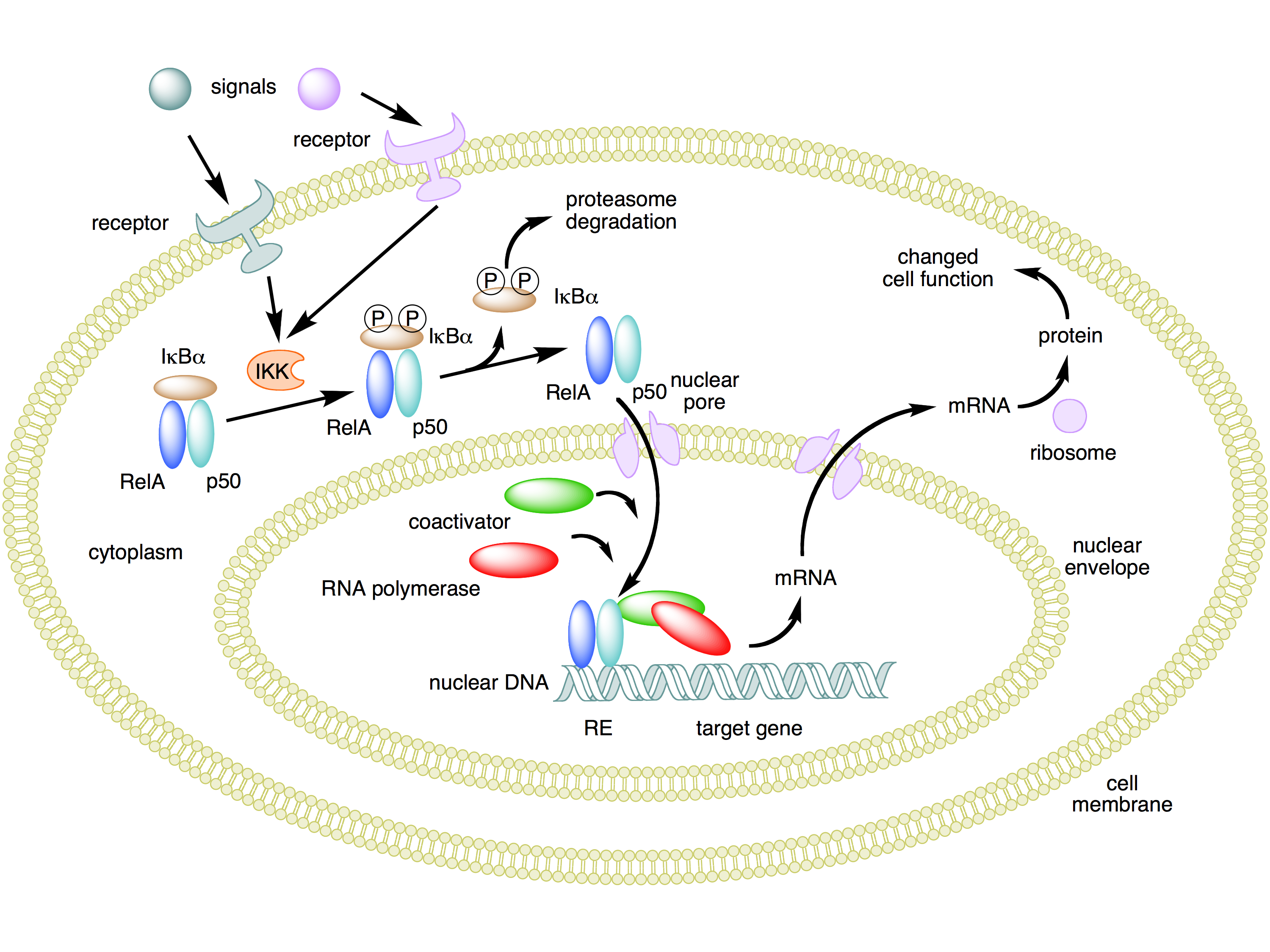
The crucial effect of inflammation for our purposes now is the fact that it activates NF-κB. It's worth noting how this process works. NF-κB is a protein complex consisting of five protein subunits, not a single protein. These subunits are RelA (also known as p65), c-Rel, RelB, p50, and p52. Complexes of these subunits normally circulate outside the cell nucleus, but they are bound to other proteins called IκBs ("inhibitors of NF-κBs") that prevent the complexes from entering the cell nucleus where they could act as transcription factors.

When an inflammatory signal binds to an appropriate cell surface receptor, signals are sent that activate kinases of a family called IKK (IκB kinase). An IKK protein phosphorylates IκBs, which in turn causes them to become unbound from NF-κB and then be destroyed by cellular proteasomes. This frees up NF-κB complexes so that they can enter the cell nucleus and affect the transcription of many genes.

The benefit of an inhibited form of NF-κB existing in the cytoplasm outside the nucleus is that it can be quickly enabled to enter the nucleus and start gene transcription when the need arises. This allows NF-κB to function as a "rapid-acting" transcription factor, without any delays caused by having to wait for the constituent proteins to be synthesized. Here's a diagram that summarizes this process. (Similar considerations apply to p53. The protein is produced and is found in the cytoplasm before it's needed, but kept from activity while bound to another protein, Mdm2.)

The next issue is what happens downstream from the inflammation-initiated activity of NF-κB in the nucleus. Since NF-κB can assist in the transcription of many different genes, the effect of its activation strongly depends on what type of cell it occurs in. Undoubtedly, there's a whole lot we don't yet know about all the affected genes and resulting downstream effects. As far as the immune response – in which NF-κB plays such an important role – is concerned, one effect involves production of cytokines for signaling to other immune system cells.

A second effect is stimulation of cell proliferation. That's generally a good thing if the cell is, for example, a B cell that mediates the part of the immune respose by manufacturing antibodies. But it can also be a bad thing when excessive proliferation of B cells leads to autoimmune diseases, leukemias, or lymphomas. NF-κB stimulates proliferation by enhancing expression of cell cycle proteins like cyclin D1.

However, there's also a third type of effect of NF-κB activity – inhibition of apoptosis. This is especially significant for cancer, because apoptosis is crucial for many natural anti-cancer cellular defenses. In particular, apoptosis is the normal response to severe, uncorrectable DNA damage. It's also the typical way that chemotherapy is able to kill tumor cells. Interference with normal apoptosis makes cancer both more likely to occur, and more difficult to treat.

Exactly how does NF-κB activity inhibit apoptosis? This has been studied, and the answer seems to be that NF-κB inhibits certain enzymes called caspases that are central to apoptosis.

There are even further suspected side effects of NF-κB activity which can play a big role in cancer. One of these is promotion of angiogenesis – production of blood vessels that can supply nutrients to solid tumors. Another is elevated expression of enzymes that promote metastasis.

Observe that the discussion here has been largely theoretical. We haven't described actual experimental research that supports the generalizations. There is some recent research already mentioned that fills this gap. It's especially interesting in the way it exposes a direct connection between NF-κB and p53. But we must leave description of that research for another article, coming very soon.

Further reading:

Nuclear factor-κB in cancer development and progression – May 2006 Nature review article

Tags: ,

Labels: , , ,

Sunday, August 16, 2009

Inflammation, microRNA, and cancer

If there's just one single point worth making about the biology of cancer, it would have to be "it's complicated".

Cells in general, and animal cells in particular, are extremely intricate Rube-Goldberg-like mechanisms. Their correct functioning depends on the integrity of 20,000 or so genes (in the case of humans), and at least 5 times as many proteins whose form is specified by the genes. Damage to even one of a few thousand important genes can put a cell on the road to becoming cancerous. So the first fact about cancer isn't really all that hard to understand: cancer (in all of its many forms) is a disease that begins with damage to the DNA of one or more genes.

This damage, which is necessary but not sufficient, can occur in many ways. Sometimes it happens because of the action of external agents, like carcinogenic chemicals or high-energy radiation (including ultraviolet light). Other times it happens simply because of occasional errors made in copying DNA during the process of cell division. These are just a few of many ways in which DNA can suffer damage. It's estimated that from 10,000 to a million DNA mutations can occur in a single human cell per day.

Fortunately, only a few percent of the 3 billion fundamental units (base pairs) of DNA actually occur within genes – everything else is "noncoding DNA". Although much of this noncoding DNA serves some useful purpose, we have little idea at present what that might be. However, it's certainly less critical to cell function than the DNA of actual genes. Even so, 10,000 or so genes in every cell could suffer mutations every day.

Of course, complex multicellular life couldn't exist unless nature had evolved some means for coping with all this random genetic damage. And so, there are a large number of ways that cells have of detecting and repairing the damage that does occur. Then in the relatively small number of cases where damage cannot be repaired, cells have additional fail-safe mechanisms to avoid malfunctions which lead to unlimited proliferation – i. e. cancer. One such mechanism is for a cell to enter a state of "senescence", where it ceases to be able to divide at all. A more drastic, but common, mechanism is for the cell to undergo "apoptosis" – orderly cell death.

A necessary condition, therefore, for a cell to become cancerous, even after DNA damage remains unrepaired (perhaps because of damage to part of the repair mechanism), is that the damage occurs in a gene that codes for proteins needed for one of the various fail-safe mechanisms. Consequently, in almost every case of cancer where a tumor has begun to form, one finds problems in some part of the cell's anti-proliferation machinery.

We'll look at a recent piece of research that identifies one particular way this can happen, and it's interesting for the variety of different cell processes that become involved.

Many of the known "causes" of cancer are fairly easy to understand. Certainly, the cancer risk from DNA-damaging carcinogenic chemicals is obvious enough. And once one understands how important a key protein known as p53 is in crucial cellular processes such as detection of unrepaired DNA damage and invocation of apoptosis if necessary, it's not hard to understand why more than 50% of human tumors have mutated genes for p53.

But there are other factors which have been found, in epidemiological studies, to be statistically associated with cancer development. One of these is inflammation, which is a very normal part of the body's immunological defenses against infection. Inflammation itself is a highly complex process – too complex to outline here. Chronic infections by various agents can cause a state of persistent inflammation. An example is the result of H. pylori bacterial infections. In addition to being responsible for stomach ulcers, such infections are also found in cases of stomach cancer. Obesity is also known as an epidemiological factor in various cancers, and the reason is now thought to be the state of chronic inflammation that obesity often causes.

What is not clear is exactly what mechanism connects inflammation with cancer. There's undoubtedly a variety of mechanisms, given how complicated cellular processes turn out to be when you get down to the finer details. The recent research mentioned above illustrated one such mechanism, in one single type of cancer.

Anti-inflammatory drugs may defeat a treatment-resistant type of cancer (6/24/09)
The research focused on a type of non-Hodgkin lymphoma called diffuse large B-cell lymphoma. In some patients with the disease, chemotherapy works well. In a recent study of 40 patients more than 75 percent of patients with one form of this type of lymphoma survived five years or longer.

But that study also identified a group of patients whose cancer proved difficult to treat. Their tumors failed to respond to chemotherapy, and only 16 percent of patients with this form of lymphoma survived more than five years after they were diagnosed.

Several molecular flags mark this treatment-resistant lymphoma, but the links between them were unknown until now. The new paper reports that tumor cells isolated from these patients have depressed levels of a protein called SHIP1, which was known to suppress tumors. In fact, patients with the lowest levels of SHIP1 are the least likely to survive.

SHIP1 is a phosphatase enzyme. That means it removes phosphate groups from proteins. So a phosphatase has the opposite effect of enzymes known as kinases, which attach phosphate groups to proteins. Having a phosphate group attached at the right place on a protein is what enables the protein to take part in a signaling pathway, which is the basic communication mechanism in a cell responsible for making things happen. Therefore, phosphatases disrupt pathways, and stop things from happening. This can be beneficial, for example, if what's happening is the excessive cell division that occurs in cancer. Accordingly, SHIP1 has been found to be a tumor suppressing protein.

In the case of diffuse large B-cell lymphoma (DLBCL), it is found that SHIP1 levels are abnormally low. It's not that the SHIP1 is defective; there's just not enough of it. So the question is why. Is there some other defective gene that's responsible?

Apparently, there is not. Instead, it's the presence of inflammation that's responsible, and in an interesting way. Inflammation is a perfectly normal product of the body's immune system, and it exists to counteract harmful agents such as bacteria. The immune system initiates and regulates the process of inflammation by means of signaling molecules called cytokines. One of the more common and important of these cytokines is TNFα.

Now, TNFα normally goes about its business without causing cancer or other lasting ill effects. In fact, under the right conditions it can induce apoptosis or inhibit tumor formation in other ways. But for some reason, in DLBCL, TNFα suppresses SHIP1, and thus promotes cancer. The research in question also discovered the mechanism of SHIP1 suppression. It turns out that the real culprit here is a small piece of microRNA called miR-155. This little bugger was already known to be involved with leukemia in mice, and with other cancers. (See references in here.)
The resistant type of lymphoma cells also have elevated levels of miR-155, a specific example of a type of genetic material called microRNA, the team found. They demonstrated that miR-155 suppresses SHIP1 by sticking to the template for the protein, preventing its manufacture. ...

The final clue came from earlier reports that an inflammatory molecule called TNFα could boost levels of miR-155. Additional laboratory work confirmed the observation for this type of lymphoma cell.

Some anti-inflammatory drugs, used for diseases such as arthritis and inflammatory bowel disease, where inflammation gets out of hand, work by suppressing TNFα. So it was hypothesized that such a drug might be beneficial in treating DLBCL. And voilà:
The anti-inflammatory drugs etanercept and infliximab, which are currently used to treat arthritis and inflammatory bowel disease, work by suppressing TNFα, suggesting a new way to curb the malignancy of this type of lymphoma.

The team tested the idea in mice that had been injected with aggressive lymphoma cells and found that nascent tumors shrank in six days.

However, mice are not humans, so the drugs need to be tested in human DLBCL patients. Patients are already being recruited for clinical studies.

Now, there are plenty of questions remaining. More needs to be understood about just what pathways SHIP1 disrupts in order to suppress tumors. This should also help in understanding why inflammation and the resulting TNFα do not, fortunately, cause cancer more often. Baby steps. But perhaps significant ones.

Here's the research abstract:

Onco-miR-155 targets SHIP1 to promote TNFα-dependent growth of B cell lymphomas
Non-coding microRNAs (miRs) are a vital component of post-transcriptional modulation of protein expression and, like coding mRNAs harbour oncogenic properties. However, the mechanisms governing miR expression and the identity of the affected transcripts remain poorly understood. Here we identify the inositol phosphatase SHIP1 as a bonafide target of the oncogenic miR-155. We demonstrate that in diffuse large B cell lymphoma (DLBCL) elevated levels of miR-155, and consequent diminished SHIP1 expression are the result of autocrine stimulation by the pro-inflammatory cytokine tumour necrosis factor alpha (TNFα). Anti-TNFα regimen such as eternacept or infliximab were sufficient to reduce miR-155 levels and restored SHIP1 expression in DLBCL cells with an accompanying reduction in cell proliferation. Furthermore, we observed a substantial decrease in tumour burden in DLBCL xenografts in response to eternacept. These findings strongly support the concept that cytokine-regulated miRs can function as a crucial link between inflammation and cancer, and illustrate the feasibility of anti-TNFα therapy as a novel and immediately accessible (co)treatment for DLBCL.


ResearchBlogging.org
Pedersen, I., Otero, D., Kao, E., Miletic, A., Hother, C., Ralfkiaer, E., Rickert, R., Gronbaek, K., & David, M. (2009). Onco-miR-155 targets SHIP1 to promote TNFα-dependent growth of B cell lymphomas EMBO Molecular Medicine, 1 (5), 288-295 DOI: 10.1002/emmm.200900028


Tags: ,

Labels: , , ,

Friday, May 02, 2008

Avian flu

Avian flu doesn't seem to be in the news so much these days, even though it continues to cause a steady stream of new human infections and often fatalities. The most heavily affected country continues to be Indonesia, but other countries that have this year reported avian flu in many domestic birds, and sometimes resulting human deaths, include Egypt, Turkey, India, Pakistan, Bangladesh, Vietnam, China, and South Korea.

An encouraging recent development is that the mechanism responsible for the serious havoc that H5N1 causes in a person's lungs is becoming better understood. It turns out to involve a signaling cascade initiated by toll-like receptor 4 (TLR-4) on the surface of macrophages in the lungs.

Macrophages are leukocytes (white blood cells) that play a role in both innate and acquired immunity. Among other things, under certain circumstances, they can cause prolific production of inflammation-causing cytokines, such as interleukin-1. When produced excessively cytokines trigger "cytokine storms" (see here), which seem to be the major harmful effect of H5N1 infection. Recently reported research gives more detail:

A single pathway for lung damage
TLR-4 is an important receptor in innate immunity, which signals activation via two pathways, TRIF and MyD88. The researchers found that lung injury was mediated exclusively through the TRIF pathway and not via MyD88. Also, the trigger for TLR-4 turned out to be the oxidation of the animals' own lung surfactant, made up of phospholipids. The oxidized phospholipids activates TLR-4, resulting in the cytokine storm that causes much of the lung damage. In order to see whether their results held true outside of the mouse-ICU, researchers tested human and simian lung samples infected with SARS, H5N1, anthrax and monkey pox and found the oxidized phospholipids in each case.

One of the interesting end results of the TRIF pathway is the activation of the NF-κB transcription factor, which in turn upregulates other proteins that stimulate T cell production, inflammation, and other additional immune response.

More: New Strategies Against Bird Flu

Tags: ,

Labels: , , ,

Sunday, April 20, 2008

Stimulating NF-κB for radiation protection

Can't resist posting about this, as it relates to one of my favorite transcription factors, NF-κB. (What' so great about NF-κB? I dunno. Maybe it's the catchy name. Besides, one of the systems it plays an important part in regulating is the immune system, especially with respect to inflammation. It's also important in the connection between inflammation and cancer. For more about it, check some other posts.)

NF-κB is actually a family of proteins, rather than a single one. It is also related to homologous proteins in a wide variety of animals, including fruit flies, sea urchins, anemones, and sponges – which is about as diverse a collection as there is in the animal department.

NF-κB is present in cells most of the time. That is, it doesn't need to await certain cell signaling events in order to be produced, which means it's always ready, albeit in an inactive state, to go to work when needed. For example, when a receptor (IL1R) for the immune system cytokine IL-1 fires up, the resulting signaling activates NF-κB that is already present.

Other receptors related to IL1R, called Toll-like receptors, can also lead to the activation of NF-κB. A specific Toll-like receptor in humans (TLR5) reacts to a bacterial protein called flagellin, which is normally found in the flagella of flagellated bacteria. That's not too surprising – this is one rather obvious way for the immune system to detect the presence of bacteria.

Now, another side of NF-κB is that it also regulates genes that control cell survival and proliferation. Specifically, it causes cells to resist apoptosis. Cancer cells take (unfair) advantage of this fact to upregulate NF-κB in order to promote their own nefarious proliferation. In particular, some cancer cells eventually become resistant to the radiation used in radiotherapy – precisely because those cells are the survivors of earlier radiotherapy.

All this background led some clever biologists to think that NF-κB could be useful in cases when one wants to protect cells from being killed by radiation – such as following a nuclear accident or explosion. It might also be helpful for astronauts on long space missions far from the Earth's protective magnetic shield against radiation.

Cancer researchers are looking for safe ways to inhibit NF-κB from protecting cancer cells. However, other biologists reasoned that doing the opposite might be a good way to protect healty cells from radiation. And further, perhaps exposing cells to flagellin might be a convenient way of activating NF-κB (instead of using an actual bacterial infection).

That line of thinking led to this:

New Drug Protects against Radiation Damage
A new drug may protect healthy tissue during cancer-killing radiation treatments or other exposures. Molecular geneticist Andrei Gudkov and colleagues report in Science this week that they protected mice from the cell-damaging effects of radiation by injecting them with a compound that helps cells resist apoptosis, or self-destruction.

Previous studies have found that cancerous cells use nuclear factor kappa-beta--a transcription factor, or protein that turns on or off a gene's protein-making ability--to outlive normal cells and grow out of control. But healthy cells in the gut switch on the same transcription factor when they interact with benign and beneficial bacteria that reside there. Specifically, the protein flagellin in some of the microorganisms' whiplike tails (which they use for propulsion) binds with a receptor on the gut cell and triggers the production of the transcription factor.

The experiment of injecting mice with flagellin and then exposing them to ordinarily lethal radiation seems to have been successful:
The injection not only protected the mice's cells but also toughened them against the effects of free radicals (molecules that can damage DNA or genetic material inside them) as well as beefed up the animal's immune systems. Mice without the injection died after the radiation treatments. "Never before has a single agent been capable of doing all three things together," [lead researcher Andrei] Gudkov says.


Further reading:

Bacteria tails could protect against 'dirty' bomb – news article in NewScientist

Drug Experiment Blocks Radiation Damage – AP news article – also here

Drug Bestows Radiation Resistance on Mice and Monkeys – news article in Science about the research.

An Agonist of Toll-Like Receptor 5 Has Radioprotective Activity in Mouse and Primate Models – technical paper in Science that reports the research.

Tags: ,

Labels: , ,

Sunday, December 23, 2007

FoxO transcription factors

Transcription factors are proteins that help regulate genes. This regulation may involve either enabling the expression of a gene or preventing expression. In the first case, the transcription factor is an "activator", and in the second case a "repressor".

Transcription factors perform their function by binding to a particular portion of DNA that is specific to a given gene. When bound to the appropriate DNA segment, a transcription factor affects gene expression by either facilitating (activator) or inhibiting (repressor) the operation of RNA polymerase in transcribing the affected gene into messenger RNA. Usually more than one transcription factor must be present to affect gene transcription, and additional proteins (called "cofactors") may also be required.

To make things even more interesting, transcription factors usually affect multiple genes, which may be otherwise unrelated to each other.

A particularly important family of related transcriptions factors comprises what are called "forkhead box" proteins, or Fox proteins, for short. (The name refers to a sequence of 80 to 100 amino acids that are part of the protein and bind to DNA, and which was originally discovered in fruit flies (Drosophila).)

Among the genes that Fox proteins are involved with are genes related to cell growth, proliferation, differentiation, longevity, and embryonic development. So there are Fox proteins that are important for things like cancer and stem cells – and thus it's quite useful to know about them.

An important subfamily of Fox proteins are the FoxO proteins, and we'll discuss some recent examples in this note.

To begin with, perhaps the most recent example is this:

Molecular Signal That Helps Muscle Regenerate Discovered (12/19/07)
Muscle regeneration after injury is complex and requires a coordinated interplay between many different processes. Key players in regeneration are muscle stem cells, so-called satellite cells. They divide and produce many new muscle cells to fix the damage incurred by injury. A crucial regulator of muscle function and repair is a signalling molecule called calcineurin. It is activated by injury and controls the activity of other key proteins involved in differentiation and the response to damage.

It turns out that calcineurin works by inhibiting FoxO.
Using sophisticated molecular techniques, the scientists revealed that calcineurin accomplishes its effect on muscle by inhibiting another protein called FoxO. FoxO is a transcription factor, a protein that plays a crucial role in skeletal muscle atrophy through the induction of genes involved in cell cycle repression and protein degradation. Suppressing the effects of FoxO, calcineurin ensures that proliferating cells stay alive and keep dividing to produce enough cells to repair muscle damage.

In this case, the normal function of FoxO is to inhibit cell proliferation (as a check on cancer), but this needs to be bypassed (temporarily) to enable muscle regeneration.

This result follows the discovery a few months earlier of the way a specific FoxO protein (FoxO1) cooperates with another important developmental protein (Notch) to control muscle cell differentiation:

Building Muscle Requires Foxo1 (8/25/07)
The mechanisms by which Foxo proteins regulate metabolism are relatively well characterized. However, little was known about the mechanisms by which these same proteins regulate cellular differentiation.

New data generated by Domenico Accili and colleagues at Columbia University, New York, now indicates that Foxo1 cooperates with Notch to control muscle cell differentiation in vitro.

Overexpression of either a constitutively active form of Foxo1 or a constitutively active form of Notch was found to inhibit the in vitro differentiation of a mouse myoblast cell line.

Note that the preceding alludes to the involvement of FoxO proteins in regulation of metabolism. This comes about because they affect the insulin signaling pathway, and hence also glucose and lipid metabolism.

This function is what allows yet another well-known protein, mTOR, to play a role in "metabolic syndrome" – a group of disorders that includes insulin resistance, heart disease and high lipid levels. (mTOR is short for "mammalian target of rapamycin". It's a protein kinase that modifies other proteins by phosphorylation.) The same mechanism appears relevant also to the "Atkins diet" and the effects of calorie restriction.

Fly Genetics Reveal Key Workings Of Atkins Diet (8/8/06)
Using fruit flies bred with a newly created mutant form of the gene TOR (short for target of rapamycin), Oldham and his colleagues were able to determine how the TOR pathway interacted with other important regulators of insulin, glucose and lipid metabolism.

TOR is an ancient gene, found in nearly all animal and plant cells. The researchers discovered that their new mutant fly reduced TOR function, allowing them to observe what happens when TOR's influence is removed.

Reductions in TOR function lowered glucose and lipid levels in the body. They also blocked the function of another important insulin regulator, a factor called FOXO, which is known to be a critical mediator of insulin signals and therefore glucose and lipid metabolism.

As if all that weren't enough, FoxO proteins are also involved with cancer and stem cells:

Gene Knockouts Reveal FoxOs' Vital Functions In Cancer Defense, Health Of Stem Cells (1/25/07)
In an elegant, multiple-gene knockout experiment, a team of Boston scientists has discovered that a trio of molecules, called FoxOs, are fundamentally critical in preventing some cancers, maintaining blood vessel stability, and in keeping blood-forming stem cells healthy. ...

The researchers at Brigham and Women's found that mice engineered to lack genes for the FoxO1, FoxO3, and FoxO4 molecules had serious blood abnormalities. Without the FoxO gene-regulating molecules, the rodents' blood stem cells -- master cells that give birth to working blood cells while also renewing themselves -- divided too fast and "burned out." ...

In the companion paper, lead author Ji-Hye Paik, PhD, of Dana-Farber and colleagues from the DePinho lab report that the three FoxO molecules, known as transcription factors, normally function as tumor suppressors that override maverick cells threatening to grow too fast and form tumors. When FoxOs are eliminated, it may allow cancer to develop.

And even that's not the end of it. FoxO proteins are also involved in the increased levels of inflammation often associated with the aging process. (This phenomenon has been tagged with the neologism "inflammaging".) It has been hypothesized that inflammaging results from the effect of phosphorylated FoxO on another notorious transcription factor, NF-κB (which is heavily involved in inflammation). Some of the effects of calorie restriction may also be due to FOXO phosphorylation. Reference: Restricting inflammaging (11/12/07)

FoxO is also regulated (as is P53) by SIRT1 – so this is yet another relationship to calorie restriction. Reference: Unlocking the Secrets of Longevity Genes

Additional references (for the seriously interested):

An AMPK-FOXO pathway mediates longevity induced by a novel method of dietary restriction in C. elegans.

Ageing: When Less Is More

Tags: , , , ,

Labels: , , , , , , , , , , , ,

Tuesday, November 06, 2007

When The Going Gets Tough, Maybe You Should Quit

This caught my eye, because of the connection with "stress", as discussed here and here:

When The Going Gets Tough, Maybe You Should Quit
Are there times when it is better to simply give up? Psychologists have been exploring this question, and more specifically a possible link between tenacity and both physical and mental health.

It would seem that persistence would be tonic over the long haul; hanging tough should increase the odds that you’ll succeed, and personal success is closely linked to well-being. But what if the goal is extremely unlikely? When does an admirable trait like perseverance start to look more like beating your head against the wall?

To test this in the laboratory, psychologists Gregory Miller and Carsten Wrosch developed a psychological instrument that can reliably distinguish between people who when faced with a difficult goal either persist or let go of it. In a series of experiments, the psychologists exhaustively studied these two personality types to see how healthy and well adjusted they are.

In their most recent study, published in the September issue of Psychological Science, a journal of the Association for Psychological Science, the psychologists followed teenagers for a full year. Over that time, individuals who did not persist obtaining hard to reach goals had much lower levels of a protein called CRP [C-reactive protein], an indicator of bodily inflammation. Inflammation has recently been linked to several serious diseases, including diabetes and heart disease.

Well, is it really surprising that there are health benefits associated with having a more easy going, "laid back" personality?

There's a very good book that delves into this in great detail – Why Zebras Don't Get Ulcers, by Stanford professor Robert Sapolsky. (Quick summary is here.)

And while we're on the subject, there's this press release that just came out:

Relationship Between Environmental Stress And Cancer Elucidated

One way environmental stress causes cancer is by reducing the activity level of an enzyme that causes cell death, researchers say.

They found that stress-inducing agents, such as oxidative stress, recruit a protein called SENP1 that cuts a regulator called SUMO1 away from the enzyme SIRT1 so its activity level drops, says Dr. Yonghua Yang, postdoctoral fellow in the laboratory of Dr. Kapil Bhalla, director of the MCG Cancer Center.

This fundamental finding about the relationship between stress and cancer opens the door for treatments that increase SENP1 activity, making it easier for cells that are becoming cancerous to die.

In yet another example of how deeply interrelated different biological processes are, it's worth noting that SIRT1 is a HDAC enzyme, whose activity seems to be enhanced by both resveratrol and calorie restriction. By mechanisms that are still somewhat mysterious, this in turn may be beneficial for longevity. If indeed oxidative stress has the effect of decreasing SIRT1 activity and hence promoting cancer, this may help explain at least some of the longevity benefit of SIRT1.

Tags: , , , ,

Labels: , , , ,

Friday, September 21, 2007

Adiponectin

As we noted in this article back in July, there are several hormones and neurotransmitters that have noteworthy effects on appetite and eating behavior, and as a result are of much interest with respect to weight gain (or loss) and obesity. The two mentioned in that article were NPY and PYY. The protein leptin is another hormone of this sort. It is often discussed in connection with obesity, because it is believed to increase metabolism and decrease appetite (as a signal of satiety). Another hormone, ghrelin, is produced in the stomach as a signal of hunger, and so it increases appetite.

A lot of research is currently appearing that deals with these substances and others, and how they are related to fat metabolism and obesity. I intend to discuss some of this research, and will begin with yet another hormone – adiponectin, which is produced exclusively in adipose tissue, i. e. fat, and hence the name. (Leptin is also produced in adipose tissue.) Unsurprisingly, adiponectin is involved in a number of metabolic processes, such as glucose regulation and the metabolism of fat for energy production.

Levels of adiponectin are inversely correlated with body mass index (BMI), and it seems to play a role in helping to stave off or ameliorate disorders such as obesity, diabetes, and atherosclerosis.

Let's begin with some older research first.

Fat Cell Hormone Causes Weight Loss (4/23/04)
Researchers at the University of Pennsylvania School of Medicine have established in an animal model that the hormone adiponectin secreted by fat tissue acts in the brain to reduce body weight. In contrast to leptin, a related hormone, adiponectin can cause weight loss by raising metabolic rate while not affecting appetite. ...

When adiponectin, which is involved in glucose and lipid metabolism, was introduced into the cerebrospinal fluid of normal mice, they showed no changes in food intake, but their metabolism rose. "The animal burns off more calories, so over time loses weight, which was very fascinating because we knew that leptin caused weight loss by suppressing appetite and increasing metabolic rate," explains [lead author Rexford] Ahima. "Here we have another fat hormone that can cause weight loss but without affecting intake."

To summarize, both leptin and adiponectin are produced in fat tissue, and both lead to increased metabolic rate. Leptin is known to be a satiety signal that also acts on the brain to decrease appetite. Although this older research suggests adiponectin has little effect on the central nervous system, we shall see that later research contradicts this.

From just about the same time we have the following, which found high levels of adiponectin in human milk, and therefore might explain the known association between breastfeeding and reduced risk of obesity later in life:

Study Detects Protein In Human Milk Linked To Reduced Risk Of Obesity (5/3/04)
The protein is adiponectin, which is secreted by fat cells and affects how the body processes sugars and lipids -- fatty substances in the blood. It's been suggested that adiponectin is involved in the metabolic syndrome, which includes insulin resistance, obesity, type 2 diabetes and coronary artery disease and occurs in 20-25 percent of adults. Higher levels of adiponectin have been associated with less disease.

If adiponectin is present in human milk, the Cincinnati Children's researchers theorized, the protein could have an influence over the metabolic "programming" of infants. That is, it could affect adiposity, or "fatness," later in life.

High levels of adiponectin were found in samples of human milk.
The researchers also confirmed the presence of leptin in human milk. Leptin is another protein produced by fat that appears to play an important role in the regulation of body fat. Leptin is a satiety hormone, involved in the state of being "full."

Adiponectin levels, however, are substantially greater than leptin in human milk, according to [lead author] Dr. [Lisa] Martin


Before we get to the latest research, here's some additional, earlier research involving adiponectin. In most of these studies, the main focus was on something else, but adiponectin was recognized as playing an important role:

Metabolic 'Footprint' May Be New Measure Of Obesity Risk In Kids (3/8/04)
Levels of a fat protein, called adiponectin, is significantly lower in overweight children and young adults. ... Adiponectin adheres to blood vessel walls, possibly protecting them by fighting inflammation at a cellular level.
Scientists Discover Obesity Disrupts Appetite Hormone, May Sabotage Body's Cues For Hunger, Fullness (7/1/04)
In addition to lower levels of ghrelin overall, the obese men showed higher levels of leptin and lower levels of adiponectin than the lean men.
Fat May Promote Inflammation, New Study Suggests (4/6/05)
In 15 study participants without diabetes, higher levels of the "bad" proteins, interleukin 6 and tumor necrosis factor alpha, were associated with a lower ability to respond to insulin and use glucose. On the other hand, higher levels of the "good" protein adiponectin were associated with an increased ability to use glucose. ... "This suggests that low production of adiponectin in subcutaneous fat is linked with an elevated risk of heart disease."
Researchers Consider Possible Mechanistic Links Between Obesity And Asthma (5/12/05)
There are also changes in the blood levels of hormones derived from fat tissue in the obese that may affect the airways. One of these hormones, leptin, is pro-inflammatory and obese individuals have higher leptin levels than lean individuals. Leptin is found at higher levels among asthmatics regardless of the extent of obesity. In contrast, blood levels of another hormone, adiponectin, which has anti-inflammatory properties, are actually lower among obese individuals.
Researchers Find Lack Of Protein In Obese People Is Risk Factor For Kidney, Heart Disease (11/28/05)
Researchers have found that mice with low levels of the protein hormone adiponectin may also have high levels of a protein called albumin which, in humans, may be a sign of kidney disease. ... To prove the relationship, they also studied mice without adiponectin (“adiponectin knockout”) compared to wild-type mice whose levels were normal. The team found that the knockout mice had three times the level of urine albumin than the wild-type mice. ... In a separate study ... researchers measured the adiponectin levels of a group of obese African American adolescents. They found similar results—subjects who had a low level of adiponectin also had the condition known as albuminuria—as indicated by high levels of the protein albumin in their urine. Albuminuria is an indicator for kidney disease.
Fat-generated Hormone Drives Energetic Capacity Of Muscle (7/6/06)
The fat-generated hormone adiponectin plays an important role in the energetic capacity of skeletal muscle, according to a new study. ... Adiponectin is unusual among fat hormones in that its levels generally decline in those who are obese. The researchers report evidence in people and mice, linking low adiponectin levels to insulin resistance and reductions in the number of "cellular power plants" called mitochondria in skeletal muscle. The findings suggest that therapies designed to boost the adiponectin signal might prove beneficial for the treatment of insulin resistance and diabetes.
New Research Could Help Women Facing High Risk Of Stillbirth (9/17/06)
They particularly looked at a key signalling molecule, mainly produced by fat cells, called adiponectin. This is known to have anti-diabetic properties as well as anti-inflammatory and anti-atherogenic actions (it prevents blood clotting which can block arteries). ... Observations showed that adiponectin levels were higher in pregnant women with type 1 diabetes at all stages of the study compared with the non-diabetic patients. Leptin levels were not different. Furthermore, they have identified adiponectin receptors on the human placenta and detected that the placenta also produces adiponectin. The researchers believe that the fetus produces adiponectin to protect itself from an adverse environment.
Weight-loss Supplement Shows Good And Bad Traits (2/1/07)
The researchers monitored insulin sensitivity in all mice throughout the study. They also monitored levels of adiponectin, a hormone secreted by fat tissue and thought to play a role in insulin resistance. “Adiponectin helps regulate insulin levels,” Belury said. “Lowered levels are associated with obesity and type 2 diabetes.” The researchers found that CLA [conjugated linoleic acid] supplementation significantly decreased body fat in the first group of mice, but at the same time excessive amounts of fat accumulated in the animals' livers. Belury and her colleagues linked this accumulation of fat in the liver to increased insulin resistance. ... But the group of mice given [insulin-sensitizer] rosiglitazone injections while on a CLA-rich diet neither lost weight nor became insulin resistant. “The drug kept adiponectin levels steady during the weeks the mice consumed CLA,” Belury said. “We think that's what kept the animals from becoming resistant to insulin.
Anti-obesity Drug May Prevent And Treat Obesity-related Liver Disease (7/4/07)
Treatment with rimonabant also normalized levels of adiponectin, a hormone that plays a key role in metabolic disorders. It is noteworthy that these results were not (or were only slightly observed) in the control animals eating the same diet but not given rimonabant, which demonstrates the beneficial effects of the drug compared to diet alone. "Our hypothesis is that the multi-protective effects of rimonabant may be mediated for a large part by both the reduction in pro-inflammatory cytokines such as TNFa and the increase in anti-inflammatory and protective cytokines or hormones such as adiponectin," the authors conclude.


In most of these earlier studies, something other than adiponectin was the main focus, yet adiponectin was recognized to have several beneficial effects, such as counteracting inflammation and insulin resistance. These effects in turn help control disorders such as diabetes, atherosclerosis, and fatty liver disease.

But let's look now at recent studies aimed at examining adiponectin itself. First off, concerning adiponectin and inflammation:

Fat Protein Cuts Blood Vessel Inflammation, May Help Heart, Scientists Find (6/24/07)
A natural substance secreted by fat cells can protect blood vessels from the damaging effects of inflammation, one of the factors that contribute to heart disease. Researchers at Jefferson Medical College have shown for the first time in an animal model that the substance – a protein called adiponectin – helps prevent immune system white blood cells from binding to the inside of blood vessel walls.

Importantly, adiponectin acted not only on leukocytes adhering to blood vessel walls, but also on inflammatory cytokines:
The scientists also looked at the effects of adiponectin on inflammation in normal mice. They gave mice a substance, TNF-alpha, which caused the release of inflammatory substances called cytokines. Injecting the mice with the active adiponectin-fragment reversed the effects of the cytokines and the resulting inflammation.

Inflammation is common in cardiovascular disease.

The next research takes a closer look at how adiponectin acts in the central nervous system:

Insulin Sensitizer Also Serves As Energy-conserving Signal To The Brain (7/12/07)
A fat-derived protein known for its effects on the liver and skeletal muscle might also serve as an energy-conserving signal to the brain during periods of starvation, suggests a new study in the July issue of Cell Metabolism, a publication of Cell Press. The substance, known as adiponectin, acts on the brain to boost appetite and slow energy expenditure in an effort to maintain adequate fat stores during lean times, the researchers report.

First off, there is the question of whether adiponectin even reaches the central nervous system.
The researchers now report evidence in mice that adiponectin receptors are present in the hypothalamic region of the brain and that some forms of the chemical enter the cerebrospinal fluid from the blood.

Then, supposing adiponectin reaches the central nervous system, there is the question of what effect, if any, it has there.
Once in the brain, adiponectin enhances the activity of a metabolic enzyme called AMP-activated protein kinase (AMPK) to stimulate greater food consumption.

Moreover, the researchers found that adiponectin decreased energy expenditure. They also showed that blood and spinal fluid adiponectin levels in the brain normally increase during fasting and decrease after refeeding, suggesting that adiponectin acts mainly during food shortages.

So this research claims that adiponectin increases appetite, unlike leptin, which has the opposite effect. Further, adiponectin leads to lower activity and energy expenditure, thus conserving available energy supplies. But such effects are reversed if adiponectin is absent:
In adiponectin-deficient mice, AMPK activity in the brain slowed, causing the animals to eat less and expend more energy. That action, in turn, made the animals resistant to becoming obese even on a high-fat diet. Moreover, animals lacking adiponectin lost more fat after 12 hours of fasting than normal mice did.

If indeed adiponectin tends to lead to lower activity levels and energy expenditure, one has to ask whether it promotes fat storage or even obesity. The next, and latest, research – which received a lot more attention outside specialist literature than research mentioned above – dramatically suggests that is the case.

The research began with mice genetically engineered to lack leptin. Without this satiety hormone, the mice overate and became quite obese. However, when a subgroup of these mice were engineered to overproduce adiponectin, they ate even more, and became almost twice as obese:

‘World's fattest mouse’ appears immune to diabetes (8/23/07)
The “world’s fattest mice”, genetically engineered to overproduce a key hormone, weigh five times as much as normal mice do – but bizarrely do not develop diabetes, reveals a new study. The findings shed light on how current diabetes medications work and point to new drug targets to treat the disease, say the study's researchers.

Philipp Scherer at the University of Texas Southwestern Medical Center in Dallas, Texas, US, and his colleagues studied mice that had been genetically engineered to overeat. The mice gorged on food because they lacked the ability to produce an important appetite-suppressing hormone called leptin.

The researchers then bred a subgroup of these leptin-deficient mice to overproduce another key hormone that gets released by fat cells, called adiponectin, by about threefold. Under normal circumstances, an increase in adiponectin levels signals that an animal has entered "starvation mode" because it has not eaten for some time.

All of the leptin-deficient mice ate non-stop, but those bred to overproduce adiponectin packed on almost twice as much weight by the end of the 20-week experiment.

Incidentally (or maybe not) it was Dr. Scherer who discovered adiponectin, in 1994.

Obviously, the most interesting outcome of this research is that the mice that overproduced adiponectin did not develop diabetes, in spite of their obesity.
Interestingly, none of the rodents that made extra adiponectin developed symptoms of diabetes, such as high blood sugar. By comparison, all of the other leptin-deficient mice developed this disease during the course of the experiment.

So why might that be?
When Scherer and his team examined the distribution of body fat within the mice, they found that the obese rodents with an abundance of adiponectin had a great deal of fat stored under the skin, but very little fat within organs such as the liver.

This unusual allocation of fat might explain why the animals remained in good health – extra fat in the liver can make the organ less sensitive to insulin, thereby leading to diabetes.

Scherer firmly believes that the distribution of fat can make all the difference in terms of whether obesity will lead to diabetes. "It's a little bit like real estate; it's location, location, location."

But wait, isn't ("type 2") diabetes mostly due to an inability to use insulin – insulin resistance? The original press release on the research ties insulin resistance directly to storage of fat in the wrong places:

Key Hormone Protects Obese Mice From Diabetes (8/28/07)
"The continual firing of adiponectin generated a 'starvation signal' from fat that says it is ready to store more energy," he said. "The mice became what may be the world's fattest mice, but they have normal fasting glucose levels and glucose tolerance.

"This indicates that the inability to appropriately expand fat mass in times of overeating may be an underlying cause of insulin resistance, diabetes and cardiovascular disease."

This discovery also suggests that in people who have low adiponectin levels fat cells don't send the signal that they're ready to accept fat, Dr. Scherer said. Instead, the fat is stored in dangerous places -- liver, heart and muscle tissues -- where it can cause inflammation and pave the way for disease.

There's at least one question left to which I don't see an obvious answer: If adiponectin is produced in fat cells ("white adipose cells", to be exact), why is it negatively correlated with obesity? That is, at least in humans, we've seen that lower levels of adiponectin go along with obesity.

That's odd. Is there some mechanism that turns off adiponectin production? Evidently so, if adiponectin normally acts as a signal of food deprivation. But exactly what is the mechanism? Would interfering with the mechanism, to keep adiponectin levels high, be worthwhile for preventing insulin resistance, inflammation, and other problems? Even if weight gain, due to increased appetite, also resulted? Needs further research, I guess.

Additional references on this research:

Tags: , , ,

Labels: , , , , , ,

Saturday, September 01, 2007

Inflammation, IL-6, NF-κB, and disease

Inflammation is a part of the body's immune response that has been implicated in a number of disease conditions, such as atherosclerosis, diabetes, osteoporosis, and cancer. Previous posts on the subject can be found here, here, here, and here.

Much of the research discussed in those posts reports on some correlation found between markers of inflammation and occurrence of disease. What we really want, however, is to understand the underlying mechanisms that might explain the correlation. Here we'll take a look at one of the hypothesized mechanisms.

This article: The Interleukin-6 inflammation pathway from cholesterol to aging – Role of statins, bisphosphonates and plant polyphenols in aging and age-related diseases is a fascinating but technical and difficult review article that makes the daring claim that
Inhibition of the signal transduction pathway for Interleukin 6 mediated inflammation is key to the prevention and treatment of aging and age-related disorders including atherosclerosis, peripheral vascular disease, coronary artery disease, osteoporosis, type 2 diabetes, dementia, Alzheimer's disease and some forms of arthritis and cancer.

Be forewarned, if you undertake to read the article, that it presumes at least a passing knowledge of biochemistry, the mammalian immune system, and the physiology behind diseases such as diabetes and cardiovascular disorders. In addition, it could be better written, with clearer development of its central arguments. Finally, its contention as quoted above is a sweeping, far-reaching hypotheses that will require much additional research to establish securely.

It is also possible that there is some element of hype in the claims made. One should always adopt a skeptical attitude towards a declaration that anything like a potential "fountain of youth" has been discovered.

This hypothesis may well be too broad. Nevertheless, the paper provides an excellent means of focusing discussion on a number of important topics that seem to be linked inevitably to strategies for preventing or delaying the onset of the aging and age-related diseases listed above. Who could fail to find that prospect interesting?

So we'll begin with some setting of the stage. Inflammation is a major feature of the mammalian immune system, which employs the vascular system (among other things) to mount a response to infections, damaged cells, and other harmful stimuli. For example, signaling proteins, known as cytokines, are dumped into the blood stream to attract the attention of other components of the immune system, such as white blood cells (leukocytes), which then migrate through the blood stream to the site of the problem.

Several cytokines play an important role in the inflammatory process. The list includes Interleukin-1 (IL-1), Tumor Necrosis Factor α (TNF-α), and Interleukin-6 (IL-6). Of these, the last, IL-6, is singled out for special attention, because it appears to be especially important in the inflammatory process itself. Control of the process is important, because research implicates an excessive or overactive inflammatory response as a significant factor in the diseases of aging listed above.

Control of IL-6, in turn, may depend on control of the protein NF-κB (Nuclear Factor κB), which is a transcription factor that is thought to be necessary for the expression of the gene for IL-6. NF-κB is an essential part of the signaling pathway through which IL-6 is produced. Without activated NF-κB there may be no IL-6. However, NF-κB is also implicated in a number of other physiological processes as seemingly independent from the inflammatory response as synaptic plasticity and memory. This poses a challenge to any attempt to regulate the inflammatory response by regulating NF-κB.

As we shall see in subsequent postings, there's a lot of very interesting research going on related to the role of inflammation in disease in general, and with the involvement of NF-κB in particular.

We'll describe here just a couple of the recent examples.

Key To Out-of-control Immune Response In Lung Injury Found
Acute Respiratory Distress Syndrome, or ARDS, is an often fatal complication of severe traumatic injury, bacterial infections, blood transfusions and overdoses of some medications. In ARDS, the lungs become swollen with fluid and breathing becomes impossible. ...

Sepsis, an overwhelming bacterial infection of the blood and organs, is the most common cause of ARDS. When the immune system responds to the infection, molecules called inflammatory cytokines and chemokines are released. These molecules attract inflammatory white blood cells and destroy bacteria, but also lead to fever, swelling and other symptoms of shock and can wreak havoc on the patient in the course of fighting off the infection.

The researchers worked with a strain of mice that lacked a gene called Cblb. This gene codes for a protein that disables a cell surface receptor. Unless this receptor is disabled it will keep NF-κB activated, and therefore leads to the overproduction of inflammatory cytokines. The result is a "cytokine storm" that leads to ARDS-like symptoms and greater likelihood of fatal results for the Cblb-deficient mice:
When sepsis was induced in mice with and without the Cblb gene, there was a marked difference in the level of the inflammatory response and survival. Mice lacking the Cblb gene were much less likely to survive than control mice.

It shouldn't be concluded, however, that NF-κB (or IL-6 for that matter) is intrinsically harmful. If that were the case, it would not be so widely conserved in evolution, as it is. NF-κB is found even in the simplest of animals, such as corals, sea anemones, and sponges.

As noted, NF-κB is a transcription factor for a number of genes besides IL-6. Some of these genes code for proteins that promote cell survival and proliferation. This, too, can be a double-edged sword, as with inflammatory cytokines. In particular, it appears that NF-κB plays a non-trivial role in various types of cancer. We'll write about that in another article. However, the following research seems to demonstrate a case where the cell survival role of NF-κB predominates:

Researchers Identify Molecular Basis Of Inflammatory Bowel Disease
[Researchers] generated a mouse model that does not express NEMO, a protein needed to activate NF-κB, in intestinal epithelial cells. As a result, these mice developed severe chronic intestinal inflammation very similar to Colitis in humans.

"A close look at the mice revealed that their gut epithelium was damaged," says Manolis Pasparakis, who recently moved from heading a lab at EMBL to becoming a professor at the University of Cologne. "NF-κB acts as a survival signal for cells. Without the molecule cells are much more likely to die and this is what happened in the intestines of our mice; individual epithelial cells died disrupting the gut lining."

Through these gaps bacteria could penetrate the intestinal wall. Right behind the gut epithelium lie cells of the intestinal immune system, the biggest immune system of our body. It detects the invading bacteria and generates a strong immune response to fight off the invaders. In the process of combating the bacteria, the immune cells secrete a cocktail of signals that bring about the symptoms of inflammation.

"This is where the vicious cycle closes," explains Markus Neurath, professor at the University of Mainz. "Inflammatory signals also reach the epithelial cells that due to the lack of NF-κB are very sensitive to them and die. The death of more epithelial cells creates bigger gaps in the gut lining so that more bacteria enter. The result is a constant immune response leading to chronic inflammation as we know it from inflammatory bowel diseases in humans."


Tags: , , , , ,

Labels: , ,

Thursday, August 23, 2007

The role of hostility, anger, and depression in inflammation

Hasn't it "always" been known that anger and hostility raises one's blood pressure? However that may be, a recent study shows that the connection of hypertension with chronic anger and hostility may involve a disturbed immune system and inflammation – in addition to the well-known blood vessel constriction that is a part of the "fight or flight" stress response.

Hostile Men Could Have Greater Risk For Heart Disease
Men who are hostile and prone to frequent intense feelings of anger and depression could be harming their immune systems and putting themselves at risk for coronary heart disease as well as related disorders like type 2 diabetes and high blood pressure, a new study finds.

The results were found in a 10-year study of U. S. veterans of the Vietnam war.
The men had a series of blood levels taken on three occasions between 1992 and 2002. Researchers measured two immune system proteins known as C3 and C4. Both are markers of inflammation, which is the body’s response to injury or infection. Changes in C3 and C4 are associated with a number of diseases, including some that negatively can affect the arteries around the heart, such as diabetes.

Men whose psychological screening showed the highest level of hostility, depressive symptoms and anger had a 7.1 percent increase in their C3 levels, while men with low levels of these attributes showed no change over the 10-year study period.

Here's another report on this research: Hostility, anger linked to chronic inflammation

But a 2004 study had already demonstrated a stronger correlation between psychological variables and a marker of inflammation (C-reactive protein):

Anger, Hostility And Depressive Symptoms Linked To High C-reactive Protein Levels
Researchers at Duke University Medical Center have discovered that otherwise healthy people who are prone to anger, hostility and mild to moderate depressive symptoms produce higher levels of a substance that promotes cardiovascular disease and stroke.

The substance, C-reactive protein (CRP), has garnered considerable attention for its role in both promoting and predicting cardiovascular disease and stroke in initially healthy people. It is produced by the liver in response to inflammation, and inflammation has recently been shown to underlie the plaque that forms inside arteries as they clog.

The Duke study is the first to link this combination of negative psychological attributes with higher levels of CRP in people without traditional risk factors for heart disease...

More specifically,
121 healthy men and women were asked to complete standard personality questionnaires in which they described their psychological attributes, including anger, hostility and depression. The volunteers did not have any pre-existing conditions -- such as smoking, high blood pressure, diabetes or heart disease -- that would predispose them to having high CRP levels. High-sensitivity blood tests were then conducted to measure CRP levels.

Respondents who were prone to anger, had high hostility levels, and showed mild to moderate symptoms of depression had two to three times higher CRP levels than their calmer counterparts. The more pronounced their negative moods, the higher CRP levels they had, the study showed.

In addition, the researcher had previously shown a relation between the psychological variables and another inflammatory substance (interleukin-6):
[H]ostile people who exhibit symptoms of depression have higher levels of stress hormones and circulating levels of an inflammatory substance called interleukin 6, another marker of inflammation that has been shown to predict heart disease in initially healthy people.

A number of other studies have demonstrated relationships between psychological stress conditions and disease states that involve the immune system, such as this one from 2006:

Anger And Hostility Speed Up Decline In Lung Power
The authors point out that hostility and anger have been associated with cardiovascular disease, death, and asthma, and that previous research has suggested that changes in mood can have short term effects on the lungs.

Anger and hostility will alter neurological and hormonal processes, which in turn may disturb immune system activity, producing chronic inflammation, suggest the authors.

An accompanying editorial comments that the physiological components of anger and stress overlap, and stress is well known to affect the immune system.

Bottom line: Get control over anger and depression if you want to stay healthy.

Of course, this is all closely related to what I discussed just a couple of weeks ago on stress and weight gain and in particular the extensive research of Robert Sapolsky summarized here.

Additional references:


Tags: inflammation, , , , , ,

Labels: , , , ,

Saturday, July 21, 2007

Health benefits of omega-3 fatty acids

Omega-3 fatty acids are now getting credit for health benefits in a surprising range of disease conditions.

Just to review a little, fats and fatty acids are said to be saturated if they have the maximum possible number of hydrogen atoms attached. In particular, a fatty acid is unsaturated if it has at least one double C-C bond on its main hydrocarbon chain. (An atom of hydrogen could potentially be attached there.) It is polyunsaturated if it has at least two. By definition, an omega-3 fatty acid is polyunsaturated, and in addition one of its double bonds occurs as close as possible to the end of the main chain that is opposite the carboxyl (COOH) group required in a fatty acid.

Curiously enough, this simple chemical property – rather than any more complicated chemical configuration – appears to be sufficient to confer a variety of health benefits on omega-3 fatty acids.

Perhaps the best-known benefit, for which there is evidence in studies of particular (not all) omega-3 fatty acids, is related to coronary heart disease (e. g. atherosclerosis or "hardening of the arteries"). The omega-3 fatty acids most frequently involved are eicosapentaenoic acid (EPA) and docosahexaenoic acid (DHA).

But recent research has encompassed many other disease conditions, for example, abnormal blood vessel growth that can cause blindness, such as that which may occur in retinopathy of premature infants, diabetic retinopathy, and "wet" age-related macular degeneration.

Omega-3 fatty acids protect eyes against retinopathy, study finds
The researchers studied the effect of the omega-3 fatty acids EPA and DHA, derived from fish, and the omega-6 fatty acid arachidonic acid on the loss of blood vessels, the re-growth of healthy vessels, and the growth of destructive abnormal vessels in a mouse model of oxygen-induced retinopathy. The retinopathy in the mouse shares many characteristics with retinopathy of prematurity (ROP) in humans. ROP is a disease of the eyes of prematurely born infants in which the retinal blood vessels increase in number and branch excessively, sometimes leading to bleeding or scarring. Infants who progress to a severe form of ROP are in danger of becoming permanently blind. There are also aspects of the disease process that may apply to diabetic retinopathy, a disease in which blood vessels swell and leak fluid or grow abnormally on the surface of the retina, and age-related macular degeneration (AMD), a disease of the macula, the part of the retina responsible for central vision, and a leading cause of vision loss in Americans 60 years of age and older.

Typical Western diets are lacking in omega-3 fatty acids, which are found mainly in shellfish and oily fish (e. g. salmon, sardines), and instead have a much higher percentage of omega-6 fatty acids. (In an omega-6 fatty acid, the only difference is that the first C-C double bond occurs farther from the end of the hydrocarbon chain that is opposite the carboxyl group. See these Wikipedia articles for more details: essential fatty acids, essential fatty acid interactions.) It turns out, oddly enough, that omega-6 fatty acids can have deleterious effects, just the opposite of omega-3 effects.
The researchers found that increasing omega-3 fatty acids and decreasing omega-6 fatty acids in the diet reduced the area of vessel loss that ultimately causes the growth of the abnormal vessels and blindness. Omega-6 fatty acid contributes to the growth of abnormal blood vessels in the retina.

To further test the apparent beneficial effect of omega-3 fatty acids, the researchers studied mice fed a diet modeled after a traditional Japanese diet (more omega-3 than omega-6 fatty acids) and mice fed a diet modeled after a traditional Western diet (lower amounts of omega-3 fatty acids). In addition, they studied mice genetically altered with a gene which mammals normally lack that converts omega-6 into omega-3 fatty acids. They found that the mice with higher amounts of omega-3 had a nearly 50 percent decrease in retinopathy.

Most importantly, this research identified a likely mechanism of action by which omega-3 fatty acids confer their benefits. The mechanism involves suppression of inflammation, especially involving the inflammatory cytokine TNF-α. In particular, this would apply to atherosclerosis, in which inflammation is generally regarded as a significant problem. Such anti-inflammatory properties, if indeed present, could account for the benefits of omega-3 fatty acids in other circumstances also. Another report on the same research describes this:

Can Blindness Be Prevented Through Diet?
Omega-3 fatty acids like DHA and EPA are thought to dampen inflammation in the body. ...

The researchers demonstrated that the omega-3-based diet suppressed production of TNF-alpha, reducing the inflammatory response in the retina, whereas the omega-6-based diet increased TNF-alpha production. The retinas of omega-3-fed mice also had increased production of the anti-inflammatory compounds neuroprotectinD1, resolvinD1 and resolvinE1. These compounds, derived from omega-3 fatty acids, also potently protected against pathological vessel growth, and they were not detected in the retinas of mice fed the omega-6 diet.


Cancer is a rather more controversial case in connection with possible health benefits of omega-3 fatty acids. Many epidemiological studies have been done to try to identify cancer-protective effects of omega-3 in the diet, with varying results. Meta-analysis of such studies does not identify a conclusive connection. However, such studies are hampered by uncertainties about the actual diets consumed by participants. Additionally, a lot may depend on individual genetic factors. In animal studies it is possible to be much more quantitatively precise. For instance, we have this:

Omega-3 Fatty Acids May Help Slow Prostate Cancer Growth
The mice were fed either a diet high in omega-3 (ratio of omega-6 to omega-3 was 1:1) a diet low in omega 3 (ratio omega-6 to omega-3 was 20:1), or a diet high in omega-6 (ratio of omega-6 to omega-3 was 40:1). The scientists compared survival rates and weighed the animals' prostates to measure tumor progression.

Mice with the tumor suppressor gene remained free of tumors and had 100 percent survival, regardless of diet. In mice with the gene defect, survival was 60 percent in animals on the high omega-3 diet, 10 percent in those on the low omega-3 diet and 0 percent in those on the high omega-6 diet.

"This suggests that if you have good genes, it may not matter too much what you eat," said [senior researcher Yong Q.] Chen, a professor of cancer biology. "But if you have a gene that makes you susceptible to prostate cancer, your diet can tip the balance. Our data demonstrate the importance of gene-diet interactions, and that genetic cancer risk can be modified favorable by omega-3 PUFA."


In a rather different direction, there has been a lot of suspicion, and some epidemiologial and experimental evidence, that omega-3 fatty acids and higher omega-3:omega-6 ratios have beneficial effects in connection with psychological and mood disorders. So it makes sense that omega-3 could be useful with the symptoms of agitation and depression associated with Alzheimer's disease. It turns out that benefits may be significantly dependent on genetic factors related to the disease:

Omega-3 Supplements Can Help With Alzheimer's Symptoms, Study Suggests
Omega-3 supplements can, in certain cases, help combat the depression and agitation symptoms associated with Alzheimer's disease, according to a clinical study conducted at the Swedish medical university Karolinska Institutet.

A number of epidemiological studies have shown that eating fatty fish provides a certain degree of protection against Alzheimer's and other dementia diseases--an effect often thought attributable to the omega-3 fatty acids it contains. Some studies also suggest that omega-3 can have a therapeutic effect on some psychiatric conditions.

The results were not straightforward, to put it mildly. There is a well-known susceptibility gene for Alzheimer's, APOE4. Carriers of the gene experienced benefits for agitation symptoms, while non-carriers had benefits for depression symptoms!
There was no observable difference in therapeutic effect between the patients receiving the omega-3 and the placebo group. However, when the researchers took into account which of the patients carried the susceptibility gene APOE4 and which did not, an appreciable difference appeared. Carriers of the gene who had received active treatment responded positively to the omega-3 as regards agitation symptoms, while non-bearers of the gene showed an improvement in depressive symptoms.


Tags: , , , ,

Labels: , ,